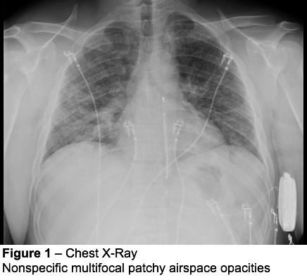
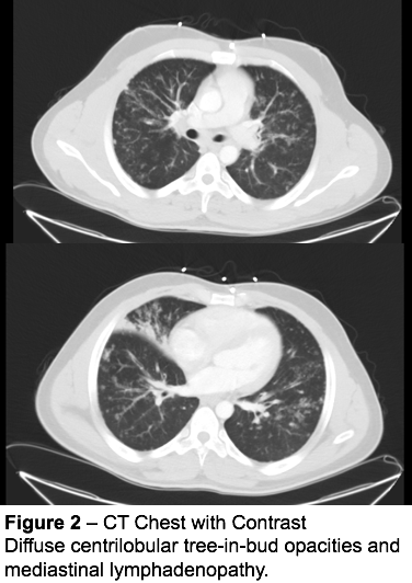
Case Presentation: A 27-year-old homeless African American man with Brugada syndrome, post automatic implantable cardioverter-defibrillator (AICD), initially presented to the hospital for acute onset of chest pain and cough for three days. Initial vitals were unremarkable; exam showed normal heart and lung sounds. EKG showed normal sinus rhythm with no signs of ischemia. AICD interrogated with no events. Labs showed troponin <0.015 (normal 0.015- 0.045), WBC 7.2 (4.5- 11.0), lactate 1.1 (0.9- 1.7 mmol/L), NT-Pro-B Natriuretic Peptide BNP 7 (0 - 450 pg/mL). He was about to be discharged when the chest X-ray resulted showing multifocal patchy airspace opacities (Figure 1).Upon further interview, patient endorsed a history of alcohol abuse, intranasal cocaine use, and vaping. He previously had been incarcerated and also was a machinist at a shipyard. CT chest scan was done (Figure 2) showing centrilobular tree-in-bud opacities and mediastinal lymphadenopathy. There was concern for tuberculosis versus pneumoconiosis/silicosis from occupational injury and the patient was admitted for bronchoscopy. During this procedure, bronchoalveolar lavage (BAL) and endobronchial biopsy was performed. AFB, fungal, bacterial cultures were negative. T-spot, Mycobacterium tuberculosis (MTB) PCR from sputum and BAL also negative. Finally, pathology from biopsy showed fibrosing non-necrotizing granulomatous pneumonitis and bronchitis consistent with sarcoid. Angiotensin converting enzyme (ACE) level was elevated at 127 (14-82 U/L).
Discussion: Pulmonary sarcoidosis is often very difficult to diagnose, especially when a patient presents with respiratory symptoms concerning for infection. In this patient, there was a broad differential on admission given his cardiac history of Brugada and nonspecific symptoms of cough. Ultimately, he fulfilled the criteria with tissue biopsy and exclusion of MTB. Interestingly, the extra-pulmonary sarcoid manifestations such as cardiac sarcoid need to be considered in this patient as well. In cardiac sarcoid, probable diagnosis includes conduction disorders (heart block, unexplained sustained ventricular tachycardia), steroid responsive cardiomyopathy, or cardiac MRI/PET findings in addition to histological extracardiac sarcoid diagnosis. This patient’s Brugada was diagnosed incidentally on EKG in a hospital admission (suicidal ideation) 5 months prior to this admission. It is unlikely that ventricular arrthymias from Brugada, which is genetic, is a component of cardiac sarcoid in this case but further investigation is needed.
Conclusions: The “tree-in-bud” sign can be commonly caused by respiratory infections including that of mycobacterial, bacterial, and viral causes. In the hospital, MTB cannot be missed. The pattern of the tree correlates to an intralobular inflammatory bronchiole and the bud correlates to inflammatory filling in alveolar ducts. Pulmonary sarcoid can show nodules in a perilymphatic distribution, but not in this particular appearance. Thus, a high degree of suspicion is required to proceed to invasive testing such as bronchoscopy, especially in a MTB rule-out. This case serves as a learning opportunity to always keep sarcoid in the differential with abnormal imaging.