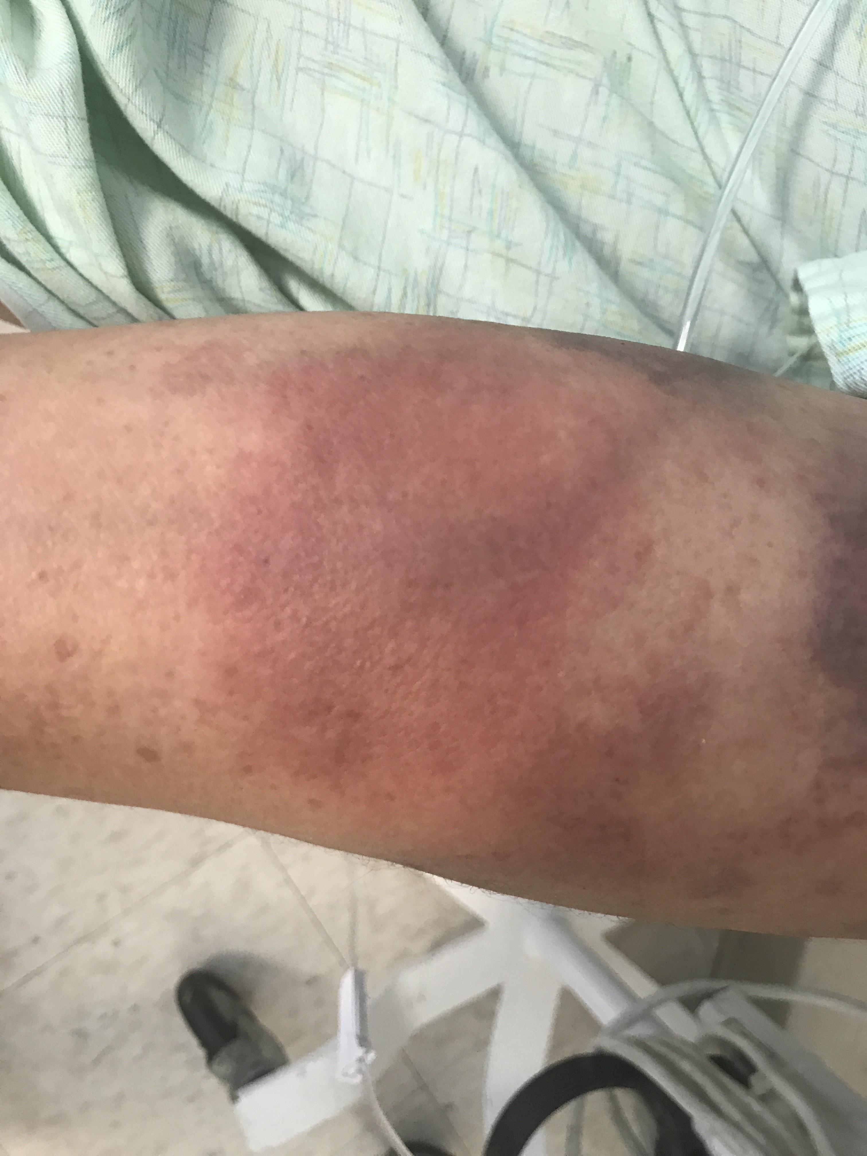
Case Presentation: A 48-year-old female with tyrosine kinase inhibitor-resistant CML on hydroxyurea therapy admitted with 3 weeks of fever, skin redness, and swelling. Her physical examination was remarkable for the temperature of 100.1 F, localized skin erythema, and tenderness around left antecubital fossa. Her labs revealed WBC of 80,000/ mm3 with 7% blast cells which dropped to the baseline level of 10,000 cells/mm3 after resuming hydroxyurea therapy. Two weeks prior to admission, she was treated with oral antibiotics for presumed cellulitis without resolution of symptoms, so we started her on broad Spectrum IV Antibiotics. Despite MRSA coverage, her symptoms persisted without any significant improvement of skin erythema or swelling. Given persistent symptoms, negative DVT study for thrombosis, and negative CT findings for necrotizing fasciitis, the skin biopsy was done to evaluate for leukemia cutis and sweets syndrome as possible etiologies Skin Biopsy showed dermis infiltration with granulocytic precursors and blast cells. Immunohistochemical stains were positive for MPO, CD15 and focal CD34, confirming a diagnosis of leukemia cutis. Bone marrow biopsy performed on the same admission and was negative for accelerated/blast phase with only 6% blast cells. She was planned for outpatient ponatinib therapy but was readmitted a week after index admission with dyspnea and tender, erythematous papular lesions on extremities. She was found to be in myeloid blast crisis as evident by WBC count of 300,000 cells/mm3 and positive flow cytometry. The patient was managed with leukapheresis and induction chemotherapy with the improvement of skin lesions.
Discussion: The patient described in this report had leukemia cutis associated with chronic stable phase of CML, preceding rapid onset (<1 week) blast crisis. Before biopsy confirmation, she was treated with antibiotics for presumed cellulitis for almost a month. Leukemia cutis is the infiltration of neoplastic leukocytes or their precursors into the epidermis or dermis resulting in clinically identifiable cutaneous lesions. It usually presents concomitantly with acute leukemias. While acute monocytic, myelomonocyte and the T cell leukemias show the highest incidence (50 to 70%) of LC, it is an uncommon manifestation of chronic myelocytic leukemia (2 to 8%). The pathogenesis of LC is not well defined but chemokine receptors and adhesion molecules have been found to play a role in skin tropism of leukemic cells. The skin lesions in LC show varied morphology; papules and nodules, indurated or hemorrhagic plaques, perifollicular acneiform papules, bullae, and palpable purpura. These lesions can be misdiagnosed as infectious and/or inflammatory cutaneous lesions and treated accordingly, as in above case. The evaluation of new skin lesions in leukemic patients should always include a skin biopsy for pathology and tissue cultures. The therapeutic management of leukemia cutis is to target the underlying malignancy.
Conclusions: Leukemia cutis associated with CML is rare. Early diagnosis is important because it is usually an impending sign of accelerated or blast phase of CML and can precede aleukaemic leukemia. New cutaneous lesions in a patient with CML should be regarded with great concern and biopsied promptly to establish the definitive diagnosis. Disease course is aggressive and early treatment with induction and consolidation chemotherapy followed by allogeneic bone marrow transplant can prolong survival.