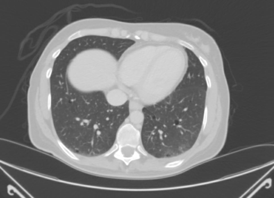
Case Presentation: Lymphangioleiomyomatosis (LAM) is a rare multisystemic disease of uncertain etiology that can have a dramatic presentation with spontaneous pneumothorax. We describe the management of a case of LAM in a healthy individual in the setting of a community hospital.A 44-year-old female with a history of smoking and vaping presented with acute onset shortness of breath. Chest radiography showed a complete left pneumothorax. A chest tube was placed with satisfactory resolution of the pneumothorax. Computed Tomography (CT) chest demonstrated scattered round thin-walled cysts throughout the lungs with basilar predominance. CT abdomen was notable only for a pancreatic cystic mass. She presented 3 days later with similar symptoms, and was found to have recurrent left sided pneumothorax on chest imaging. Robot-assisted thoracic surgery (RATS) with mechanical pleurodesis & wedge biopsy of the lung was performed. Biopsy pathology revealed smooth muscle proliferation, establishing the diagnosis of LAM. A rheumatologic workup was negative. Follow-up pulmonary function testing (PFT) was normal, and the decision was made to monitor disease progress with frequent PFTs.
Discussion: LAM is frequently found in association with tuberous sclerosis; sporadic LAM is a rare entity, with an incidence of 3 to 5 per million women (1). The precise pathophysiology is uncertain, but is thought to involve excessive proliferation of atypical smooth muscle cells due to aberrant stimulation by estrogen and other growth factors. Clinical manifestations include fatigue, dyspnea, and spontaneous pneumothorax. Extrapulmonary manifestations include renal angiomyolipomas, uterine and cerebral lesions, along with lymphatic manifestations including chylothorax and chyloperitoneum. A characteristic feature of LAM is the presence of diffuse, bilateral, thin-walled pulmonary cysts, the formation of which has been speculated to be due to smooth muscle cell proliferation within the airways with consequent distension of terminal airspaces. A workup for LAM should include the measurement of VEGF, the elevation has a sensitivity and specificity of 71% and 100% respectively for diagnosis (2). Abdominal imaging should also be pursued to delineate the extent of visceral involvement. Our patient had minimal symptoms prior to presentation, and also had normal PFTs, requiring a lung biopsy to reach a diagnosis. Treatment options include sirolimus and everolimus, which are mTOR kinase inhibtors, with management of pneumothorax with mechanical or chemical pleurodesis. LAM is a rare cause of spontaneous pneumothorax, and should be considered in a middle aged woman with diffuse cystic lung disease and pneumothorax with no other clear causes, even with normal lung function testing.
Conclusions: Hospitalists should consider LAM as a part of the differential diagnosis when encountering a patient with a spontaneous pneumothorax, especially when cystic lung disease is noted.