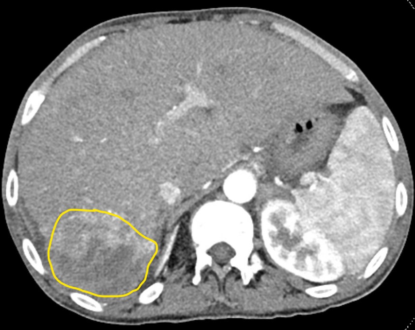
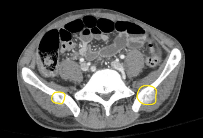
Case Presentation: A 33 year old male with a history of hypoplastic left heart (treated by staged surgeries during infancy including the Fontan procedure at age 3), autism spectrum disorder, and intellectual/developmental disability presented to the emergency department (ED) with progressively worsening lower back pain that started 5 days prior to presentation. An outpatient screening liver ultrasound obtained a week prior showed cirrhotic morphology with two hepatic lesions measuring up to 3.1cm. A CT of the abdomen was then performed a day prior to presentation which showed enhancing hepatic lesions measuring up to 9.1cm (Picture 1). Because of this imaging, he was referred to the ED where his vitals were only remarkable for heart rate of 107. His BMP was unremarkable and CBC only significant for a Hgb of 12.0. Liver function test (LFT) was significant for alkaline phosphatase of 223 and an INR of 1.4. Alpha fetoprotein (AFP) was significantly elevated to 7256. An MRI of total spine and a CT pelvis were then obtained. These showed an enhancing lesion at L3 vertebral body with resulting severe canal stenosis. There were also other enhancing lesions at C2, multiple thoracic vertebrae and ribs, bilateral iliac wings (Picture 2), and the sacrum. He was then admitted to General Medicine, specifically our hospital’s med-peds team that cares for young adults with chronic childhood-onset conditions (CCOC). During admission, neurosurgery was consulted with no acute surgical interventions recommended. A CT-guided biopsy of the left iliac wing was performed. The patient was discharged home once his postoperative pain was controlled. Medical oncology and radiation oncology were consulted and scheduled immediate followup. The pathology later confirmed poorly differentiated hepatocellular carcinoma (HCC). After discussion with the oncology specialists, the patient’s mother opted to focus on palliative radiation and pain management.
Discussion: Liver disease and HCC are known complications of patients who have received the Fontan procedure as the purpose of the procedure is to change the hemodynamic flow to the heart and lungs(1). This in turn causes increased hepatic venous congestion, which can lead to Fontan-associated liver disease (FALD), cirrhosis, and eventually can progress to HCC. The incidence of cirrhosis and HCC is highest in patients 20 years or more out from Fontan(2). The mean age of diagnosis of HCC associated with FALD is 30 years(3), which is much younger than HCC in the general population. Because of the higher risk of FALD and HCC in these patients, international guidelines recommend LFTs and AFP at least every 1-2 years for adults who received the Fontan procedure and frequent liver ultrasounds every 6 months if there is any concern for cirrhosis(4). Unfortunately, this patient was unable to get frequent surveillance due to transportation difficulties, which likely led to a delayed diagnosis. The prognosis of patients with Fontan-associated HCC is poor with survival rates of 57% and 35% at 1 and 5 years, respectively(1).
Conclusions: At many institutions, adult survivors of congenital heart disease are cared for on cardiology teams. However, as the number of these patients continue to increase, it is likely that more will be seen on hospitalist teams, especially for those that care for young adults with COCC. While the risk of FALD and HCC in Fontan patients may be well known to congenital cardiologists, it may not be as commonly known among hospitalists and should be a can’t-miss diagnosis in this population.