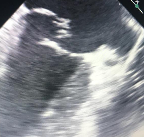
Case Presentation: A 68-year-old woman with a history of hypertension and possible interstitial lung disease presented with one week of worsening midsternal chest pain, diaphoresis and nausea. Initial vitals were unremarkable. Exam revealed bibasilar crackles but no murmur, gallop, elevated jugular venous pressure or abdominal tenderness. An electrocardiogram showed no acute ischemic changes. Initial troponin I was 3.23ng/mL. Complete blood count and comprehensive metabolic panel were unremarkable except for 12,600 white blood cells per microliter. The patient was given aspirin 325mg and a heparin drip was started. She was taken for urgent coronary angiography which revealed no obstructive disease. However, troponins continued to increase over the next two days to a peak of 8.26ng/mL. Computed tomography showed no evidence of pulmonary embolism but showed bilateral hilar adenopathy and fibrotic changes suspicious for atypical distribution of sarcoidosis. Cardiac magnetic resonance imaging showed a normal ejection fraction, no wall motion abnormalities, no prior myocardial damage, no evidence of sarcoidosis and normal mitral valve function. The etiology remained unclear and then five days after presentation the patient developed a grade IV/VI systolic murmur best heard at the apex. Echocardiogram showed new severe mitral regurgitation, and the patient was taken for emergent mitral valve replacement. Pathology demonstrated acute inflammation and necrosis of the underlying myocardium.
Discussion: Patients frequently present with chest pain and elevated troponins in the absence of acute ischemic changes on electrocardiogram. Based on risk factors, hemodynamic stability and lab abnormalities, certain patients should be taken for urgent coronary angiography. Despite non-obstructive findings on coronary angiography, patients can still have acute myocardial infarction. Papillary muscle rupture is one of the many known complications of myocardial infarction and can have deadly consequences. However, patients infrequently present with papillary muscle rupture after myocardial infarction with non-obstructive coronary artery disease.
Conclusions: Myocardial infarction may initially result in only symptoms and cardiac enzymes abnormalities without ischemic changes on electrocardiogram or blockages visible on coronary angiography. Coronary angiography and other types of imaging modalities can be useful in the diagnosis of myocardial infarction but have their limitations. Physicians must maintain a high degree of suspicion for potentially deadly sequelae of myocardial infarction such as papillary muscle rupture even without clear angiographic evidence of myocardial infarction.