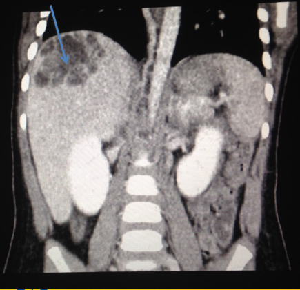
Case Presentation: A 3-year-old previously healthy male was admitted with a five-day history of fevers up to 104 F, decreased oral intake, and decreased activity. He had been seen by his pediatrician on day three of illness, at which time he was noted to have leukocytosis with a white blood cell count of 30,200/µL and erythrocyte sedimentation rate of 37 mm/hr. His symptoms did not improve with antipyretics. He had decreased oral intake but his review of systems was otherwise negative. He did not have any known exposure to infectious agents or any recurrent infections.On presentation to our facility, his physical exam was significant for mild right upper quadrant abdominal tenderness and hepatosplenomegaly. His laboratory evaluation was significant for elevated inflammatory markers, hypoalbuminemia, and elevated liver enzymes without any eosinophilia. His throat, urine, and blood cultures as well as his viral respiratory panel were all negative. An abdominal ultrasound showed a mass in the periphery of the right hepatic lobe. Computed tomography of the abdomen confirmed the presence of a hepatic cystic mass and a small right pleural effusion. He was started on empiric Metronidazole, Vancomycin, and Ampicillin/Sulbactam. Echinococcus antibody, Entamoeba histolytica antibody, Entamoeba histolytica enzyme immunoassay, and parasitic antibodies were sent but were negative. The mass was not drained immediately due to concern for anaphylaxis if it was indeed a hydatid cyst.Despite antibiotic therapy, he remained febrile, and his hospital course was complicated by the development of a new left pleural effusion which required placement of chest tubes. He eventually had a drain placed in the liver abscess, and the fluid culture was positive for Klebsiella pneumoniae. Vancomycin and Metronidazole were discontinued and he was continued on a three-week course of Ampicillin/Sulbactam. Serial abdominal ultrasounds were performed which showed eventual resolution of the abscess. He also had resolution of his pleural effusion as well as his other clinical symptoms.
Discussion: The differential diagnosis for a hepatic mass is broad and includes liver abscess, neoplasm, hemangioma, and hydatid cyst, among others. Multiple bacterial organisms, including Escherichia coli, Klebsiella pneumoniae, and Pseudomonas aeruginosa have been implicated in the development of pyogenic liver abscesses. In pediatric patients, the incidence is 10 cases per 100,000 hospital admissions although it is more common in people of Southeast Asian descent. Risk factors for the development of pyogenic liver abscesses include diabetes mellitus and immunocompromised state. The most common route of bacterial spread is through the biliary tract and portal circulation. Continuous spread from an adjacent source of infection, systemic bacteremia, and hematogenous spread are also possible. Associated infections include meningitis, brain abscesses, and endophthalmitis. Common presenting symptoms include abdominal pain, vomiting, fevers, and chills. In more severe cases, patients may develop altered mental status and shock. Management of pyogenic liver abscesses generally requires a combination of antibiotics and percutaneous or surgical drainage.
Conclusions: Since the presenting symptoms may be non-specific, timely diagnosis may be difficult. Klebsiella pneumoniae liver abscesses can be an unusual source of persistent fevers and may present a diagnostic challenge for healthcare providers.