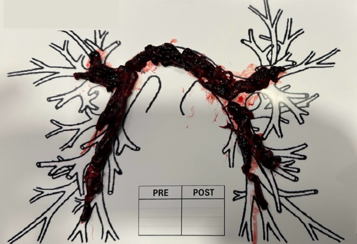
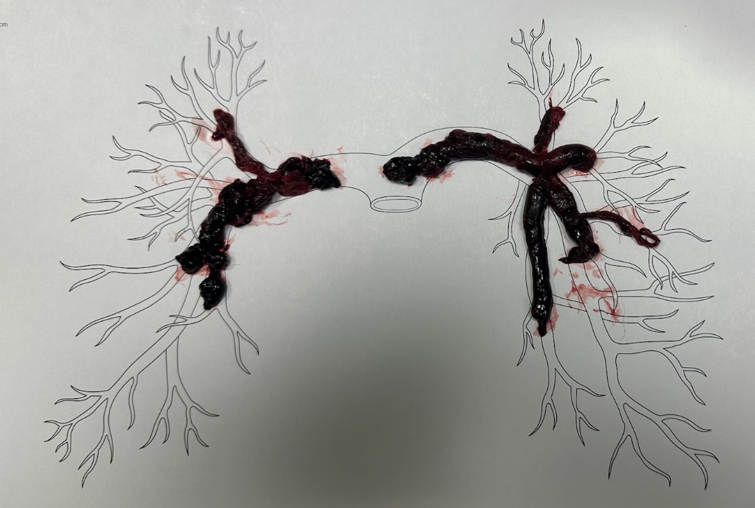
Case Presentation: A 38-year-old woman with no significant past medical history, no current medications, and no history of smoking presented with worsening dyspnea one month after foot surgery, which had been marked by prolonged postoperative immobility. On arrival, CT pulmonary angiography revealed a large saddle pulmonary embolism (PE) with extensive bilateral lower lobe thrombi and right ventricular strain. Interventional radiology performed a suction thrombectomy, successfully removing extensive thrombi and reducing pulmonary arterial pressure from approximately 70 mmHg to 20 mmHg.In the MICU, the patient developed worsening confusion and hypoxemia, prompting repeat imaging that revealed a recurrent saddle PE with associated pulmonary infarction. She underwent a second suction thrombectomy with improvement in pulmonary hypertension, and an infrarenal IVC filter was placed. Despite initial stabilization, she subsequently required intubation and mechanical ventilation with intermittent proning for 72 hours due to concern for reperfusion injury and ARDS. Additionally, she had an inadequate response to unfractionated heparin, prompting transition to enoxaparin and later a direct oral anticoagulant.A comprehensive thrombophilia evaluation was negative. She gradually improved, was extubated, and discharged in stable condition.
Discussion: This case illustrates the complexity of evaluating a severe PE in a patient whose risk factors do not fully account for the magnitude of disease. Although our patient had recent surgery, immobility, and obesity all likely contributing to a provoked VTE, the severity of bilateral DVTs and a submassive saddle PE at age 38 was atypical. A thrombophilia workup was clinically justified, and although negative, her clot burden remained out of proportion to her identifiable risk factors.The patient’s early clinical deterioration following the initial thrombectomy underscores the need for monitoring after intervention. Persistent thrombus burden shortly after the initial procedure reinforces that clot fragmentation or thrombus propagation can occur, requiring prompt reassessment and re-intervention. Her progression to ARDS reflects a recognized complication of reperfusion, associated with sudden restoration of blood flow and alveolar-capillary disruption.Additionally, her difficulty achieving therapeutic anticoagulation on heparin introduced another layer of complexity. Heparin resistance can occur in the setting of elevated factor VIII levels, inflammatory states, or antithrombin III deficiency. Transitioning to low molecular weight heparin allowed for more reliable anticoagulation during a period of increased thrombotic risk.
Conclusions: This case demonstrates the importance of considering underlying hypercoagulable states in atypically severe VTE and recognizing complications such as residual thrombus, reperfusion injury, and anticoagulation resistance. Ultimately, timely multidisciplinary intervention and ongoing reassessment facilitated a successful recovery.