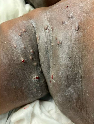
Case Presentation: A 56-year-old female with history of HTN presented to a community hospital campus of our system with three days of fever, headache, nasal congestion, and myalgias. Her temperature was 100.8 F and HR 102 BPM; labs showed WBC of 13.2 x 10 E/uL, and chest X-ray revealed a hazy posterior lower chest opacity. She was admitted for community-acquired pneumonia and treated with IV antibiotics and fluids. She remained febrile and in the hospital for the next week when she developed a tender, pruritic, umbilicated papulonodular rash on the breasts, inframammary folds, groin, and labia. Studies recommended by Rheumatology consultants showed elevated CRP of 296.8 MG/L and ESR of 120 MM/Hr with RF of 16 U/mL and negative ANA and ANCA titers. Workup per Infectious Disease consultants was negative for HSV, VZV, MPox, EBV, HTLV1, TB and HIV. She was transferred to our campus for dermatology evaluation and a punch biopsy of the rash. A CT chest revealed a left breast nodular density without any residual pneumonia. One day after transfer, the rash became ulcerative with purulent exudate. After ulceration, the rash became more concerning for infection, vasculitis, paraneoplastic process, and Sweet syndrome. General surgery was consulted and determined the breast lesion was unlikely to be cancer and could be followed up in the outpatient setting. The punch biopsy revealed neutrophilic dermatosis consistent with Sweet syndrome. High-dose corticosteroids were initiated, leading to rapid defervescence within 48 hours and rash resolution over the following week without recurrence.
Discussion: Sweet syndrome is an inflammatory skin disorder classified into three types: classical, malignancy-associated, and drug-induced. Classical Sweet syndrome is often related to an infection of the lungs or GI tract and is the most common type. We present here a case of Classical Sweet syndrome after a pneumonia. This case was complicated by the breast lesion found on CT scan which initially raised concern for malignancy but was eventually ruled out in conjunction with our specialists. Given the historical association between Sweet syndrome and malignancy, availability and anchoring bias may have contributed to the initial focus on a malignant trigger. Our patient developed a rash one week after upper respiratory symptoms, consistent with the typical 1 to 3-week latency between prodromal symptoms and rash onset. Hypersensitivity to foreign antigens and cytokine dysregulation are thought to mediate the development of Classical Sweet syndrome. Drug-induced Sweet syndrome was unlikely, as the patient had no exposure to granulocyte-colony stimulating factor, immune checkpoint inhibitors, or high-risk antibiotics such as trimethoprim-sulfamethoxazole and nitrofurantoin, which usually cause reactions within two weeks of exposure. Given the benign morphology of the breast density, and the exclusion of high-risk medications, the presentation was most consistent with classical, post-infectious Sweet syndrome.
Conclusions: This case highlights that early recognition and treatment of Sweet syndrome can rapidly improve symptoms in patients with unexplained inflammatory skin disease. Clinicians should include Sweet syndrome in the differential for post-infectious or drug-induced rashes. Awareness of its multiple triggers may reduce anchoring on malignancy as the presumed cause.