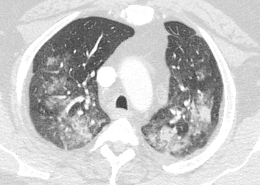
Case Presentation: A 50-year-old male with history of gastric lap band, obstructive sleep apnea, hypertension, and type 2 diabetes presented with two months of worsening shortness of breath. He denied cough, fever, myalgias, diarrhea, loss of sense of taste or smell, or pleuritic chest pain. He had no history of asthma, COPD, or tobacco use. He was hospitalized for acute hypoxic respiratory failure requiring non-invasive positive pressure ventilation. CT chest showed bilateral multifocal ground glass opacities more peripherally located typical of COVID-19 pneumonia. Dexamethasone was started. COVID-19 was negative via nasal swab on 2 separate occasions. RVP was negative. Bronchioalveolar lavage (BAL) was also negative for COVID-19. Cefepime and vancomycin, were started for suspected sepsis secondary to bacterial multifocal community acquired pneumonia. However, the patient developed worsening hypoxic respiratory failure requiring intubation. He was treated presumptively for Aspergillus with voriconazole, which was stopped after multiple negative tests. He had no risk factors for recurrent aspiration. No home medications were associated with drug induced hypersensitivity pneumonitis. Infectious work-up of sputum culture, BAL culture, blood cultures, fungal blood cultures, serum galactomannan, serum 1,3-beta-D-glucan, Legionella urine antigen, Blastomyces antibody, Coccidioides antibody, and histoplasma antibody were all negative. Autoimmune workup had a speckled ANA of <1_80 which was nonspecific and negative anti-DS DNA, ANCA, anti-Jo-1, anti-smith, anti-RNP, CCP, rheumatoid factor, anti-SCL-70. BAL cytology was without malignant cells. Alpha-1 antitrypsin was negative. Transthoracic echo was normal. The patient slowly improved without a clear explanation and was extubated. Rheumatology and infectious disease were consulted and unable to find a definitive etiology. After a two month hospitalization, he was discharged home with the working diagnosis of cryptogenic organizing pneumonia.
Discussion: Although COVID-19 pneumonia has a characteristic appearance on CT chest with peripherally located ground glass opacities (1), this is not pathognomonic, and a careful history should always be attained. In this case, the history of two months of progressively worsening subacute dyspnea without viral upper respiratory tract symptoms was inconsistent with COVID-19 pneumonia, prompting a more thorough work up. Sensitivity of nasal swab for COVID-19 was 78.3% in one study (2) while BAL around 93% (2). Our patient had three negative nasal swabs for COVID NAA followed by a bronchoscopy with BAL which was also negative, effectively ruling out COVID-19 pneumonia. A differential diagnosis for subacute hypoxic respiratory failure should include fungal pulmonary infections, autoimmune diseases such, interstitial lung diseases, recurrent aspiration pneumonitis, and drug associated pneumonitis. Although yet to be diagnosed the most likely culprit given the lack of improvement with antibiotics and antifungals, completely negative autoimmune workup, no concerning home medications, lack of chronic aspiration, is interstitial lung disease. 10-15% of all ILD cases are considered unclassifiable and never given a definite diagnosis (3).
Conclusions: When the clinical history does not correlate, hospitalists should maintain a broad differential for diseases that may mimic the symptoms and imaging of COVID-19, including bacterial and fungal pneumonias, autoimmune disorders and ILD.