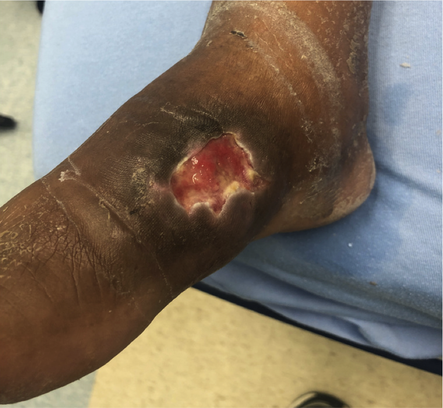
Case Presentation: 73 year-old Eritrean woman presented with one-month history of non-healing foot wound. While visiting Eritrea, she developed right foot edema, leading to splitting of the skin on the dorsal foot. This developed into a wound requiring hospitalization, debridement, and two week antibiotic course with ampicillin and ciprofloxacin in an Eritrean hospital. No organism was identified. She returned to the US three weeks later with enlargement of the wound, which never seemed to heal. She had no fever, respiratory symptoms, or swelling of the leg. She denied fresh or salt-water exposure or close livestock exposure, but occasionally wore open sandals. On exam, she was well appearing with no lymphadenopathy, normal lung exam, with an ulcer of right mid-foot with a clean pink base and slough tissue present at the margins without drainage or erythema (picture 1). Differential included fungal or bacterial wound, mycobacterial infection, including buruli ulcer, sarcoid lesion, autoimmune dermatologic ulcer, rare tropical disease, including eumycetoma, yaws, or cutaneous leishmaniasis. Laboratory analysis revealed negative HIV Ab and RPR. MRI showed soft tissue ulceration with osteomyelitis of 1st and 2nd metatarsal, medial cuneiform, medial navicular bones. Operative debridement was performed. Intraoperative appearance of the wound was characterized by soft tissue destruction and bone necrosis with paucity of purulence; the surgeon commented it looked as though bone was disintegrating. Smear microscopy of surgical pathology was negative for acid-fast bacilli. Aerobic wound broth culture grew coagulase-negative staphylococci, however, this was not a satisfying diagnosis for prolonged non-healing ulcer. During this time, Interferon‐Gamma Release Assay resulted positive. Pulmonary tuberculosis was deemed unlikely given absence of respiratory symptoms, normal chest CT imaging, and negative sputum MTB nucleic acid amplification test (NAAT). This further raised suspicion for extra-pulmonary TB (EPTB) or non-tuberculosis mycobacteria. Twenty-three days after surgery, intraoperative broth culture grew Mycobacterium tuberculosis complex with good susceptibility. Vancomycin was discontinued and she was placed on RIPE therapy for 2 months.
Discussion: EPTB represents around 15-30% of all forms of TB (1,2). Skin and soft tissue M. tuberculosis infection is among the more rare clinical manifestations comprising approximately only 1 to 2% of all TB cases (3,4), although bone and joint infection is more common. In this case, absence of foot pain prior to development of the soft tissue wound suggests directly inoculated cutaneous infection leadinge to osteomyelitis. Scrofuloderma, contiguous spread to the overlying skin from adjacent structures, such as bone, is another possibility. Well-organized epithelioid granulomas, which we suspect in this case, are often associated with a high degree of cell-mediated immunity (CMI) and low bacillary load, which may complicate diagnosis.
Conclusions: The diagnosis of cutaneous TB is challenging due to varied clinical presentations, shared histopathology with other disease, and often low bacillary load. Although sensitivity for DNA PCR for diagnosis of EPTB varies, studies reporting sensitivity from 4.5 – 88% (5,6), use of NAAT on tissue may have expedited diagnosis and antimicrobial sensitivity. Inquiring about the sensitivity of a lab’s NAAT for an EPTB suspicious lesion may aid in more rapid diagnosis and initiation of antitubercular therapy.