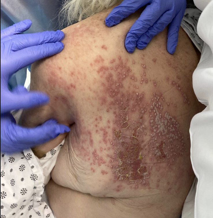
Case Presentation: An 88-year-old woman with diabetes mellitus and dementia initially presented with symptomatic bradycardia with loss of consciousness. Her hospital course was extended while awaiting nursing home placement. On hospital day (HD) 18, the patient complained of burning back pain. The exam revealed an erythematous, confluent vesicular rash confined unilaterally to the T2-T4 dermatomes on her left back. A presumptive clinical diagnosis of herpes zoster was made and oral valacyclovir was initiated. Daily examination of the rash showed an increasing number of vesicles on an erythematous base with crusting and scaling [Figure 1]. On HD 24, there were new vesicles crossing the midline with involvement of bilateral axillae. A singular lesion was seen on the upper lip along with mucositis. Infectious disease (ID) and dermatology were consulted and agreed with the diagnosis of herpes zoster and treatment with oral valacyclovir. Varicella zoster PCR and IgM antibody were negative. On HD 28, a punch biopsy of the rash was performed. Due to clinical presentation concerning for dissemination and lack of improvement on oral valacyclovir, treatment was switched to intravenous valacyclovir. A Tzanck smear was negative for multinucleated cells. The final dermatopathology result of the punch biopsy revealed tinea corporis. The patient was treated with topical ketoconazole with a reduction in the vesicular and crusting appearance of the rash without further spread and improved pain.
Discussion: Tinea corporis is a fungal infection caused by dermatophytes that affect the skin of the trunk, neck, arms, and legs. It commonly presents as an annular, erythematous, and pruritic rash, but can mimic other dermatoses [1]. Factors that affect the morphology of atypical tinea presentations include the pathogenicity of the specific dermatophyte species and host factors such as immune status, underlying comorbidities, and mechanical insults [2, 3]. In our case, tinea corporis was initially diagnosed and treated as herpes zoster. The case emphasizes the importance of being mindful of anchoring and confirmation biases, especially when treatment does not yield expected improvement. When considering alternative differential diagnoses of dermatoses in hospitalized patients, along with serological studies, skin scraping microscopy with KOH preparation and a skin biopsy should be performed, especially if initial less invasive diagnostic testing is negative [4].
Conclusions: Dermatophytosis can present in an atypical form due to both patient and organism factors. In cases of presumed herpes zoster that do not improve with empiric treatment, it is imperative to consider tinea corporis in the differential and perform appropriate testing.