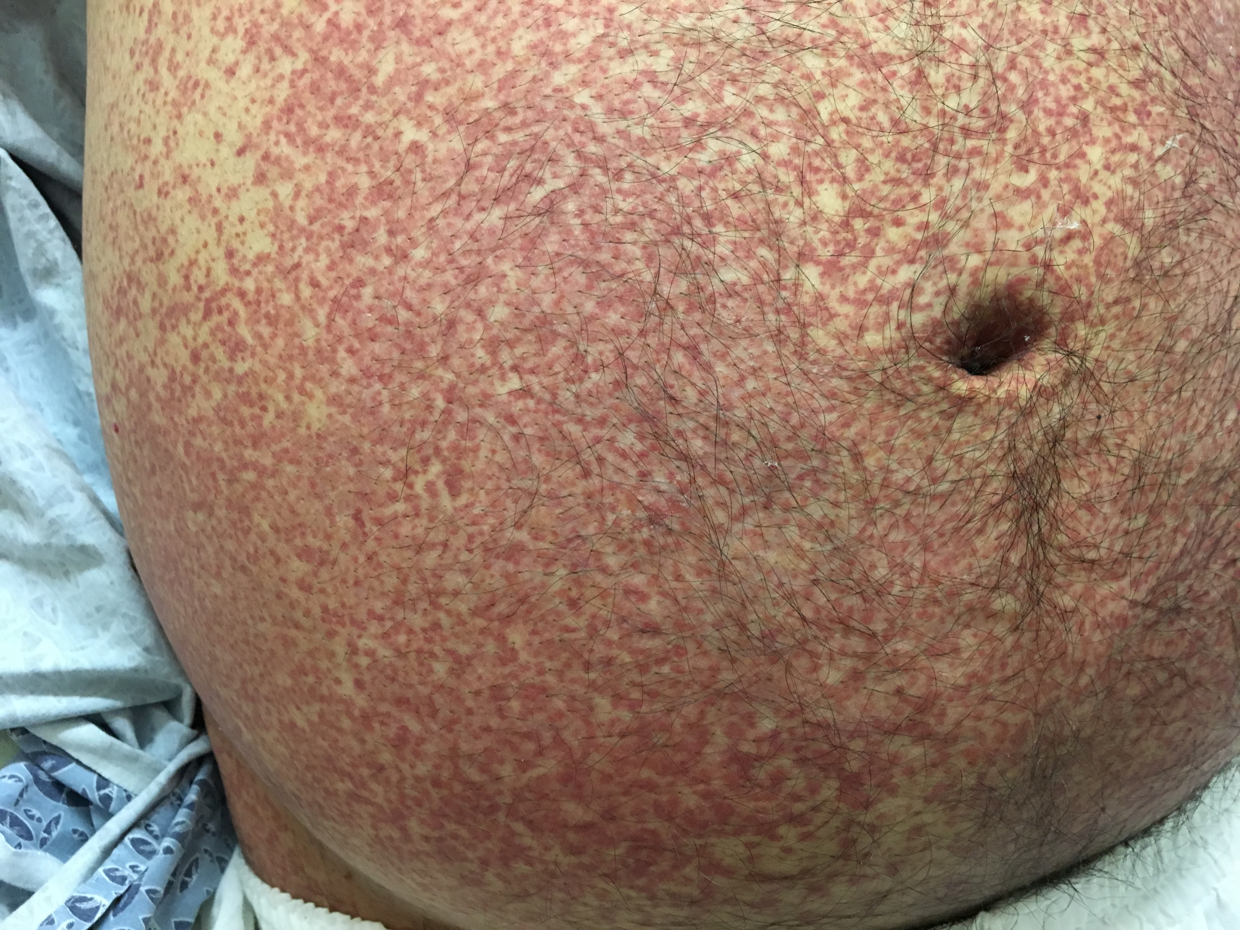
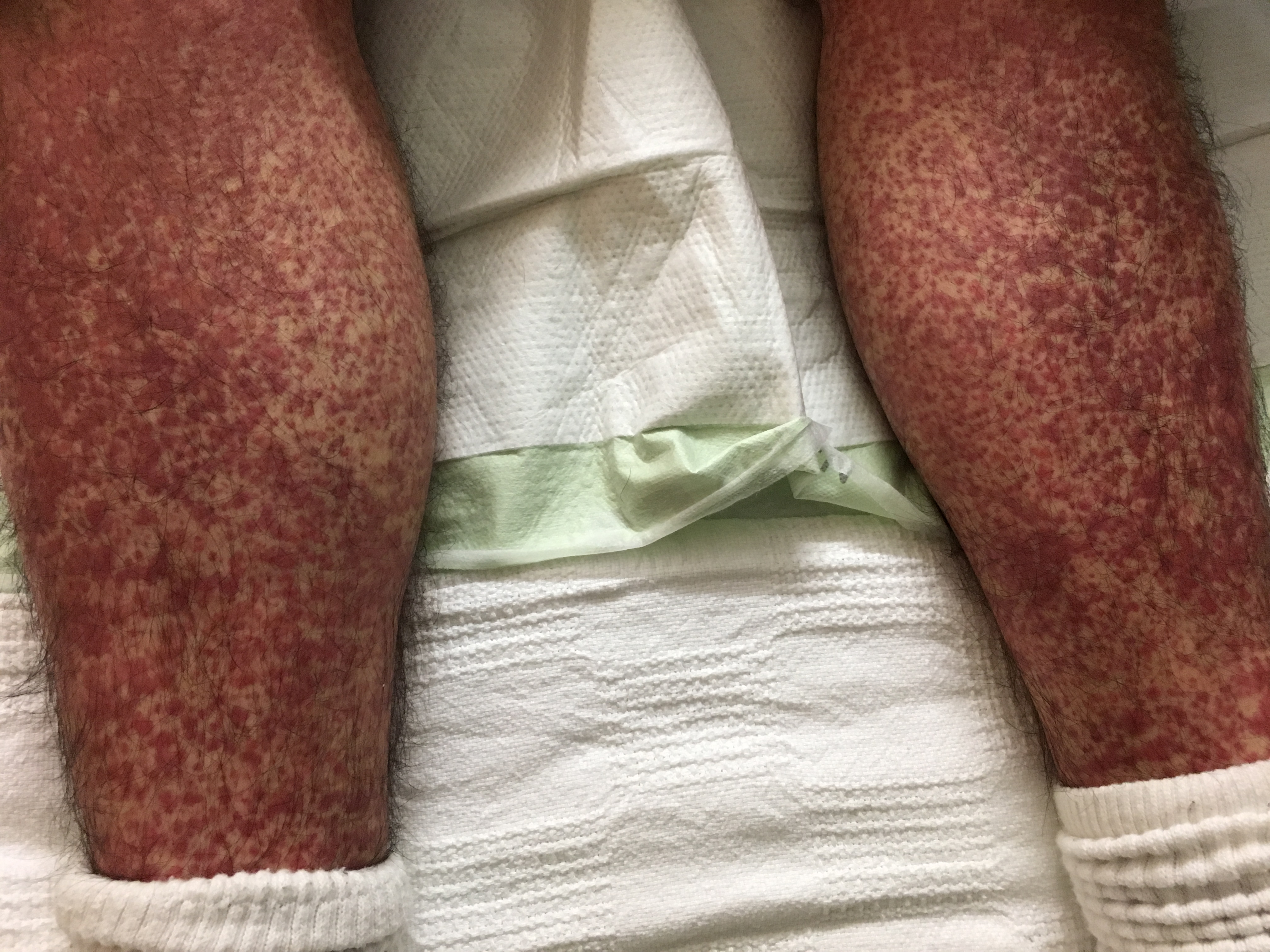
Case Presentation: A 36-year-old man presented with 10 days of progressive rash that started on his abdomen. The lesions grew, developed a more intense violaceous color, and spread to his lower back, buttocks, forearms and lower legs. The rash spared his upper torso, upper arms, neck, face and palmar/plantar surfaces. He also reported chills, nausea, vomiting and fatigue. He denied taking any medications and had no sick contacts, recent travel or insect bites. Vital signs were unremarkable except for a heart rate of 104. Physical exam revealed a right eye conjunctival hemorrhage and subtle abdominal discomfort to palpation without organomegaly. Integumentary exam demonstrated innumerable palpable purpura that were non-blanching, with some lesions coalescing. There were no lesions in the oral mucosa. Pertinent laboratory data revealed normal renal function and mild thrombocytopenia. A urinalysis showed microscopic hematuria and trace proteinuria. HIV, HCV, HBV, anti-nuclear and anti-neutrophil cytoplasmic antibody testing were all negative. CRP and ESR were both elevated. Complement levels were normal. Dermatology performed a punch biopsy. Pathologic analysis revealed superficial and mid-dermal perivascular infiltrate composed of neutrophils and neutrophilic debris as well as extravasated red blood cells. The samples stained strongly under immunofluorescence for immunoglobulin A (IgA) and C3, confirming the diagnosis of IgA vasculitis. While awaiting biopsy results, the patient developed severe colicky upper abdominal pain and recurrent vomiting. Corticosteroids were initiated, and he had considerable improvement in his skin rash and abdominal symptoms. A repeat urinalysis showed no hematuria. The patient was discharged home to continue prednisone and follow-up with his primary physician and dermatology.
Discussion: Many skin lesions are harbingers of systemic illness and can have significant morbidity and mortality if a diagnosis is incorrect or delayed. In this case, a small-vessel vasculitis emerged as the suspected culprit, and a biopsy confirmed an IgA-associated leukocytoclastic vasculitis. IgA vasculitis is the most common systemic vasculitis in children; however, the disease can occur in adults. Most cases are idiopathic, but other causes include infections, medications or autoimmune conditions. Palpable purpura is the most common manifestation, but involvement of joints, gastrointestinal tract and kidneys are possible. Bowel ischemia and renal failure are life-threatening complications. Though debate exists on treatment with steroids or other immunosuppressive agents, most cases have a benign course and resolve spontaneously.
Conclusions: Acute-onset skin disorders are often a diagnostic challenge for hospitalists, and we are frequently tasked with the initial assessment and management of these diseases. With prompt evaluation and consultant involvement, a diagnosis of IgA vasculitis can be quickly established, and treatment can yield rapid recovery in both the patient’s rash and potential end-organ manifestations.