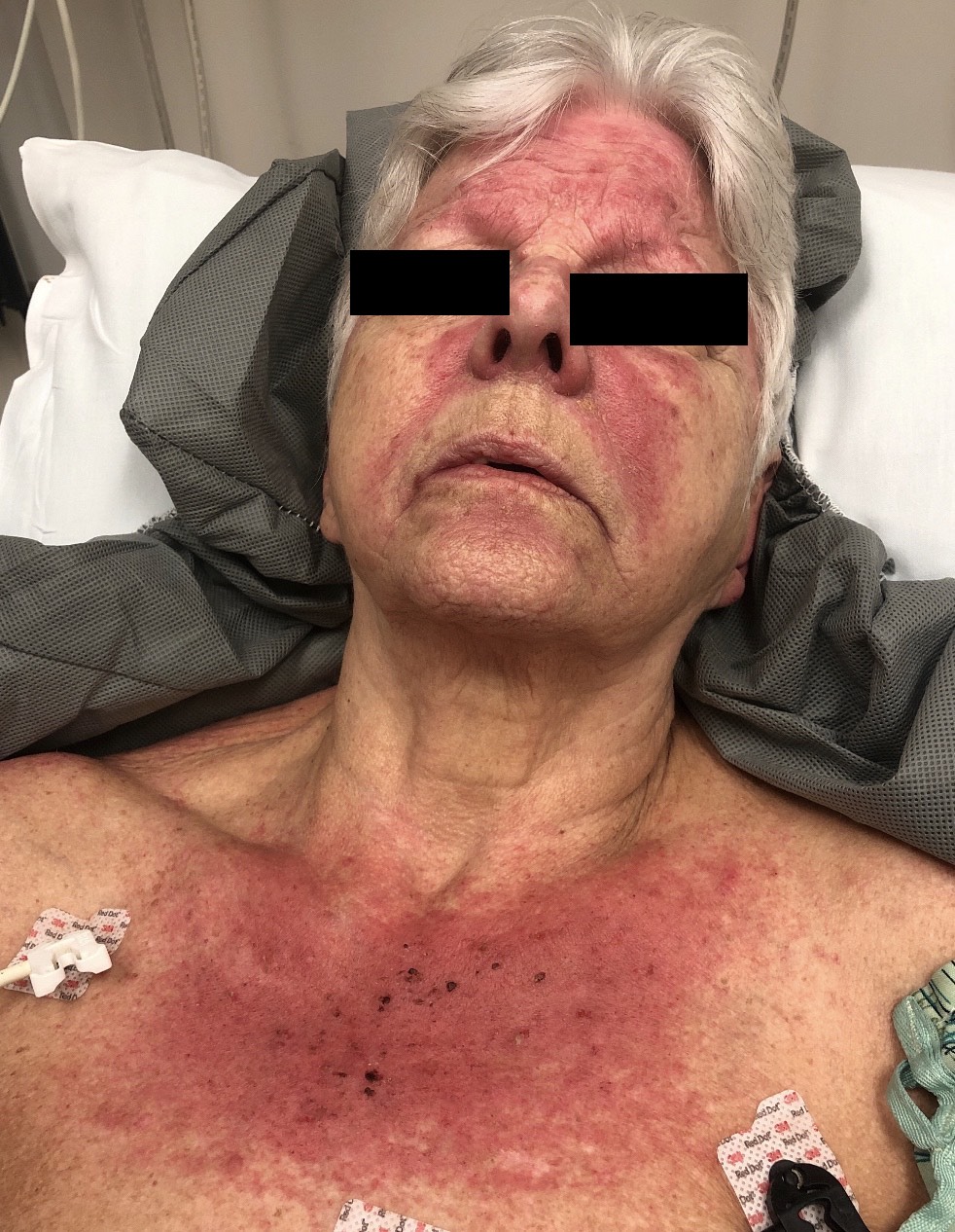
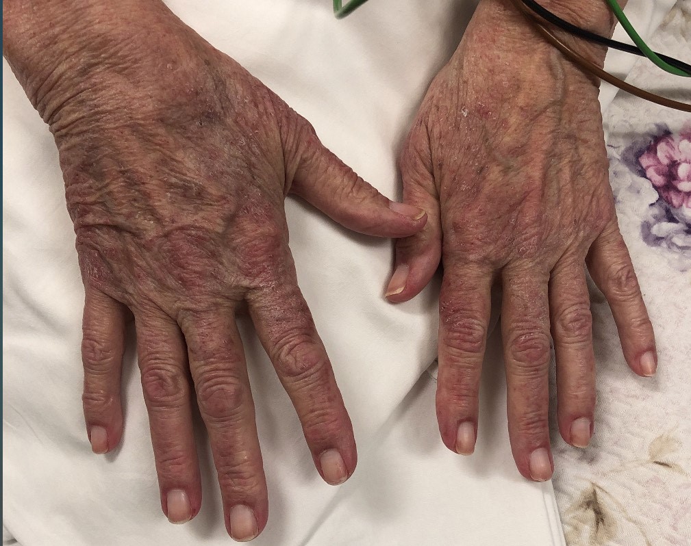
Case Presentation: The patient is a 77-year-old woman who presented with a two-day history of progressively worsening oropharyngeal dysphagia, dysphonia and left upper extremity weakness. She was discharged from the hospital three days prior for a three-month history of a pruritic rash. During the previous hospitalization, she was noted to have facial erythema, Shawl and V signs, and Gottron’s papules. She was evaluated by dermatology and underwent a skin biopsy with a preliminary pathology consistent with dermatomyositis. She had also mentioned that she was experiencing some dysphagia, which she attributed to oral mucosal ulcerations, and generalized myalgias and fatigue, which she attributed to a recent prolonged hospitalization. Despite these subjective findings as well as elevated creatine phosphokinase and C-reactive protein levels, she was discharged only on topical steroids with a presumed diagnosis of amyopathic dermatitis. After discharge the patient experienced worsening of her rash, and had profound worsening of her dysphagia, being unable to swallow both liquids and solids, prompting her to return to the hospital. During her second hospitalization, she was noted to have 3/5 left upper extremity strength, as well as pharyngeal collapse and glottic gap on laryngoscopy. She was evaluated by dermatology and rheumatology, both of whom had agreed upon a formal diagnosis of dermatomyositis. She was initiated on systemic steroids with significant improvement in her symptoms.
Discussion: Dysphagia and rashes are common symptoms that are encountered by hospitalists. Dermatomyositis is a type of idiopathic inflammatory myopathy and can present with a wide range of symptoms, most notably proximal muscle weakness, cutaneous findings, interstitial lung disease, dysphagia, and polyarthritis. A diagnosis is usually made using the EULAR/ACR classification criteria in conjunction with physical examination findings. Treatment for dermatomyositis involves high dose systemic glucocorticoid therapy followed by a slow taper, and likely an addition of a glucocorticoid sparing agent. There is a subset of individuals that are diagnosed with amyopathic dermatomyositis when only cutaneous manifestations are present. These patients can, at times, be treated with topical steroids, photoprotection and anti-pruritic agents, but most also need and benefit from systemic steroids.
Conclusions: This patient’s elevated muscle enzymes and inflammatory marker, in addition to presence of dysphagia and generalized myalgia should likely have prompted evaluation by rheumatology to help distinguish dermatomyositis versus amyopathic dermatomyositis. Although it is impossible to predict, given the patient’s significant response to systemic steroids, treating the patient with steroids during and after her first hospitalization may have prevented her readmission.