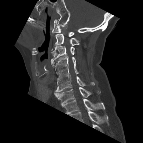
Case Presentation: A 70-year-old female with past medical history of cervical (C4/C5/C6) spine fusion presented with one-year history of swallowing difficulties. She noted that over the past year she has developed steadily worsening swallow ability from solids to liquids associated with frequent coughing when eating. She has also lost approximately 20 pounds due to reduced swallow ability. On physical examination, her gross motor and sensory functions were normal, and no pathologic reflex was detected. EGD showed esophagitis, erosive gastritis, and no strictures or masses. Further study, with a barium swallow, revealed a normal oral phase and a severely impaired pharyngeal phase. She had significant posterior pharyngeal wall edema at C2-C4, limiting the passage of bolus and causing significant pharyngeal residue that subsequently was aspirated. Swallow was severely impaired secondary to a large osteophyte. CT cervical spine showed prominent osteophytes from the level of C2-C4 indenting the posterior glottis. ENT and Neurosurgery was consulted; the patient underwent an anterior cervical spine osteophyte C2-C3 reduction. Patient swallowing improved after the surgery, and during the two-month follow-up, patient stated that her swallowing was back to normal.
Discussion: Cervical osteophytes are common and often asymptomatic in the aging population. Causes of cervical osteophytes include conditions such as diffuse idiopathic skeletal hyperostosis, ankylosing spondylitis, degenerative changes, prior trauma, or following spinal surgery. Rarely do cervical osteophytes grow anteriorly, but when it occurs, they can cause disruptive symptoms including dysphagia, dysphonia, and dyspnea. Dysphagia related to cervical osteophytes can mimic esophageal malignancy as it appears progressively, initially with solids and then with fluids. Physicians should be aware of this presentation, especially in the elderly population. The mechanisms to explain the symptomatology include mechanical compression and inflammation secondary to local mass effect. Diagnosis is made with a CT scan defining the bony anatomy and a barium swallow to confirm the presence of esophageal compression by the cervical osteophyte. Treatment usually depends on the symptoms. Management for patients with slow progressing disease includes diet measures with soft and semiliquid foods. Patients with laryngeal edema are given antireflux medication and steroidal anti-inflammatory medications. In cases like ours where dysphagia is severe and approaches aphagia causing substantial weight loss, surgery is the best option. Depending on their size and their projection, the osteophytes can usually be resected using an osteotome. The risks of surgery include hematoma, esophageal perforation, infection, and resection or compression of nerves around the anatomical area. After surgery, most patients report improvement of the symptoms. Food intake can be resumed after recovery from anesthesia and patient passes a swallow evaluation.
Conclusions: Dysphagia is the sensation of food being caught during the passage from the mouth through the esophagus into the stomach. It’s classified into oropharyngeal or esophageal etiologies. Most common causes of esophageal dysphagia include esophageal dysmotility, inflammatory conditions, and structural abnormalities. Cervical hyperostosis causing a mechanical compression and inflammation of the esophageal lumen is recognized as an unusual cause of dysphagia.