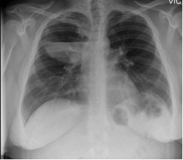
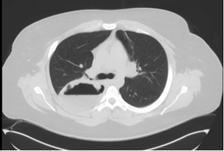
Case Presentation: A 23-year-old male with intermittent controlled asthma, presented to the emergency department for recurrent hemoptysis and night sweats of two months duration. The patient also complained of loss of sensation in his left thumb, index and middle fingers and bilateral lower extremity rash of two days duration. On examination, lung fields were clear to auscultation bilaterally with normal percussion. Neurologic examination was remarkable for decreased tactile sensation in left hand of ulnar distribution. Skin examination was remarkable for palpable purpuric lesions bilaterally on the lower extremities. Chest X-ray revealed a large cavitary lesion with air fluid level in the right upper lobe. Computed tomography of the chest further defined the cavitary lesion size as 11.5cm*7cm with significant fluid and irregular walls with extensive pleural contact. Laboratory results were remarkable for white blood count of 19 thousand/ul with 42% eosinophils (7.960 thousand/uI). Autoimmune work-up revealed positive proteinase 3 anti-neutrophil cytoplasmic antibody (c-ANCA PR3+). A punch biopsy of the purpuric rash performed before initiation of steroids revealed leukocytoclastic vasculitis with a large number of eosinophils in capillaries and post-capillary venules. After multidisciplinary discussions involving pulmonary, rheumatology, and infectious disease consultants, a diagnosis of polyangiitis overlap syndrome was made. Patient was treated with pulse dose steroids with clinical improvement and was discharged with maintenance therapy of Rituximab.
Discussion: A cavitary lung lesion has a wide differential and includes infection, septic emboli, malignancy, and non-infectious granulomas. Less common etiologies include sarcoidosis, pulmonary infarct and cryptogenic organizing pneumonia. Given this broad differential, it is important to determine the underlying etiology and treat it appropriately. In our case, patient had several features that pointed towards the diagnosis of vasculitis, in particular small vessel vasculitis. The diagnosis of the underlying vasculitis was challenging as patient had features overlapping granulomatosis with polyangiitis (GPA) and eosinophilic granulomatosis with polyangiitis (EGPA). After discussion with the consultants, our patient was diagnosed with polyangiitis overlap syndrome, given his history of asthma, peripheral and tissue eosinophilia which are more associated with EGPA and presence of PR3 positive C-ANCA and cavitary lesions which are more consistent with GPA. Of the 15 cases of GPA- EGPA polyangiitis overlap syndrome reported in literature, 4 cases described nodular lesions on chest CT, 9 cases reported ground glass opacities and 2 cases reported cavitary lesions. Therefore, Polyangiitis overlap syndrome needs to be considered as a part of differential diagnosis while evaluating patients with cavitary lung lesion. Our patient was appropriately treated with methylprednisolone 1mg/kg for 3 days inducing a decrease in peripheral and tissue eosinophils. Patient’s symptoms improved and he was discharged on Rituximab for immunosuppression. During post hospitalization follow up, he was noted to have significant reduction in size of his cavitary lesion.
Conclusions: Cavitary lung lesions are a common presentation of several disease entities and vasculitis needs to be considered as a part of differential diagnosis. It is also important to identify the type of vasculitis as treatment differs significantly.