Case Presentation: A 36-year-old female presented with a month history of cramping abdominal pain, nausea and vomiting that started after an episode of diarrheal illness associated to ingestion of mangoes. Her symptoms were worse with food intake and consequently, she reported a 25-pound weight loss. Her PCP had performed a urea breath test and an abdominal ultrasound, both of which were unremarkable. She had been taking over the counter NSAIDs without improvement. On admission, she was afebrile and hemodynamically stable. Her physical exam was remarkable for diffuse tenderness to palpation, but no peritoneal signs were present. All laboratories, including liver function profile and lipase were normal. Due to marked weight loss, she underwent upper endoscopy, which was unremarkable. CT abdomen and pelvis showed a high-grade partial small bowel obstruction with transition point in the right lower quadrant. Nasogastric tube was placed for decompression and surgery was consulted. She underwent exploratory laparotomy and was found to have dense adhesions and scarring of the terminal ileum and cecum and apparent ileal stricture. Ileocecectomy and ileocolonic anastomosis were performed. Pathology of surgical specimens, including terminal ileum, cecum and ascending colon showed severe endometriosis with transmural involvement and serosal fibrous adhesions associated with stricture formation in terminal ileum. The patient recovered well after the surgery.
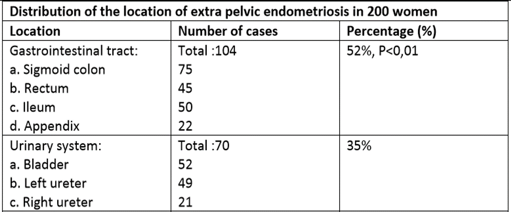
Discussion: Endometriosis affects up to 15% of women of reproductive age. Classic symptoms include dysmenorrhea and infertility, however, only 25% of affected women present with these classic symptoms, making the diagnosis challenging. When the bowel is involved, patients can present with diarrhea, constipation and bleeding. These symptoms can be initially related to menstrual cycles; however, they can become permanent as the lesions progress. Although colonic involvement is common, the incidence of small bowel involvement is only 0.5%. Several imaging modalities have been proposed to aid in preoperative diagnosis including transvaginal ultrasound and CT enterography, which commonly shows constrictive bowel lesions that can be easily mistaken for inflammatory bowel disease. Therefore, the gold standard for diagnosis continues to be laparoscopy. In cases of complicated bowel endometriosis, like our patient, this is also the preferred therapeutic approach.
Conclusions: Given the diagnostic difficulties, it is important to keep a high index of suspicion for bowel endometriosis in patients of childbearing age who present with non-specific gastrointestinal symptoms.