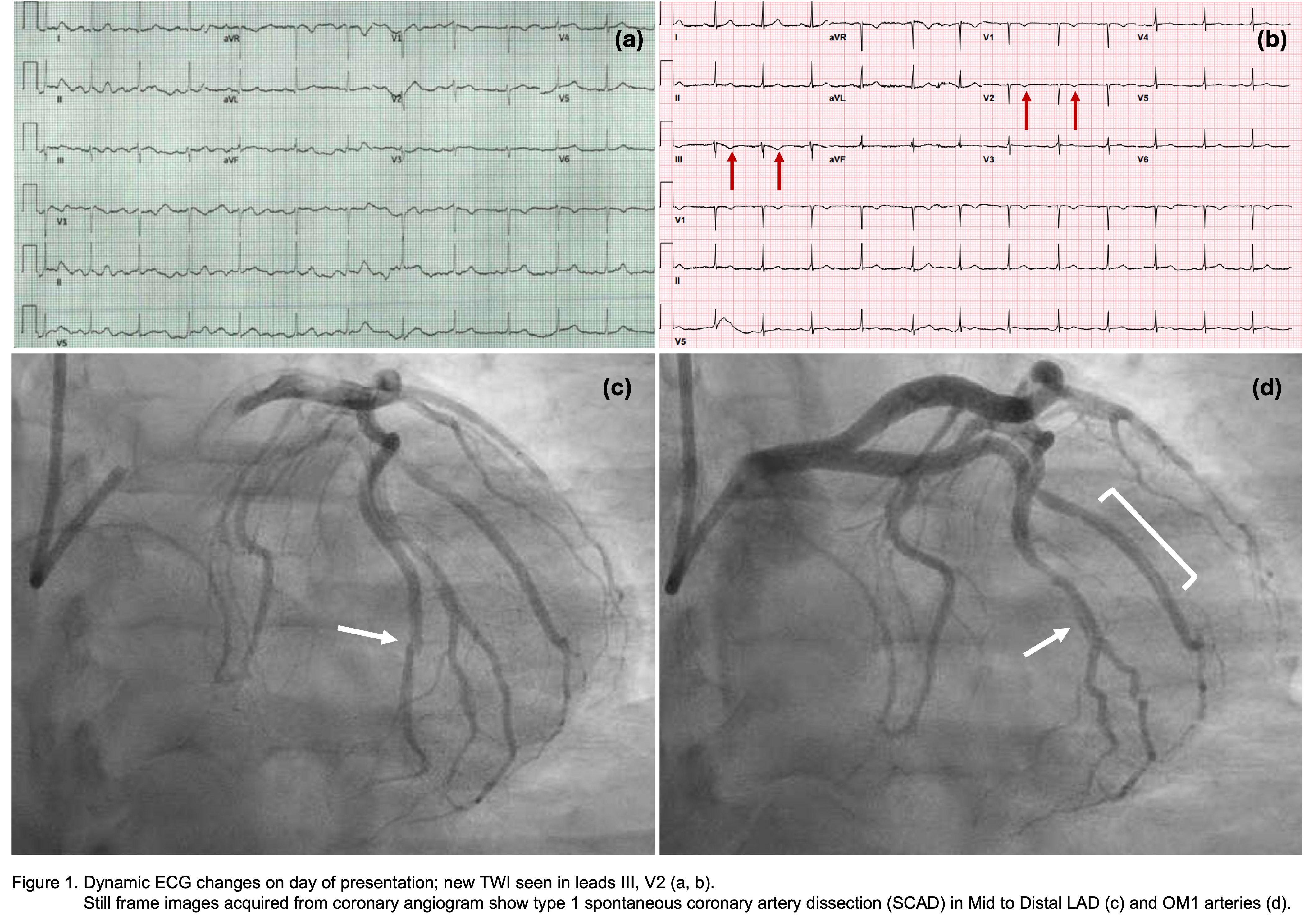
Case Presentation: A 30-year-old G2P1A1 woman with a history of gestational diabetes, pre-eclampsia, hypertension, and recent medical termination of pregnancy at around 8 weeks with mifepristone and misoprostol presented to the ED for pressure-like, non-radiating midsternal chest discomfort and dyspnea. Blood pressure was 135/105. Family history included MI in her grandfather at age 50-60, and heart failure in her father at age 70. She denied drug use or smoking. Initial EKG showed sinus rhythm with nonspecific ST-T changes and poor R-wave progression (Figure). High-sensitivity troponin was 32, rising to 5,392. Bedside ultrasound showed preserved EF with no obvious regional wall motion abnormalities. Repeat EKGs showed T-wave inversions in leads III and V2, suggesting dynamic changes. Cardiac catheterization (Figure) showed type I SCAD in the mid-distal LAD and LCx-OM1, with TIMI 3 flow in the distal vessel. The patient was treated medically with metoprolol succinate, statin, 1 month of dual antiplatelet therapy (aspirin and clopidogrel), followed by 11 months of aspirin alone. TTE showed preserved LVEF 60% and hypokinetic apical and mid-lateral walls. The patient was counselled to avoid future pregnancy. Fibromuscular dysplasia (FMD) was considered; MRA abdomen showed a questionable irregularity of the left renal artery (but no beading) and incidentally, median arcuate ligament syndrome (MALS). During follow up, the patient had two ED visits for chest pain without evidence of ischemia. Referrals were made to FMD and cardiovascular genetics clinics.
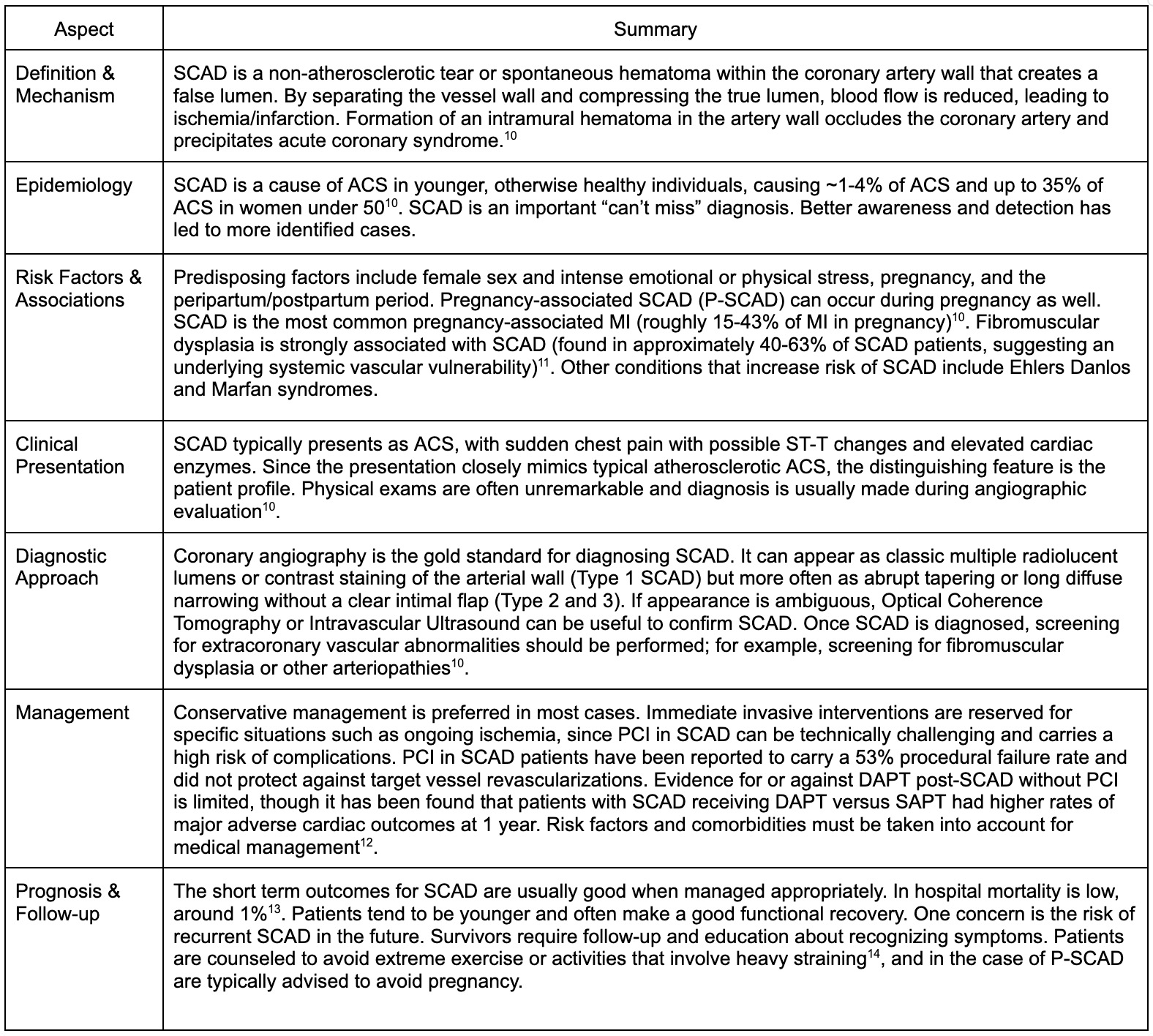
Discussion: Spontaneous coronary artery dissection (SCAD) results from separation of coronary arterial wall layers, due to intimal tearing or spontaneous intramural hematoma [1]; it causes up to 25-30% of myocardial infarctions (MI) in women under 50 [1] and has been linked to pregnancy, stress, and arteriopathies such as FMD [1]. This case highlights the need to be vigilant for SCAD in young women with angina, particularly near pregnancy. While pregnancy-associated SCAD (P-SCAD) is well described and is the leading cause of pregnancy-related MI [2], SCAD post pregnancy termination is unusual, with sparse reports of SCAD after abortion, stillbirth, or misoprostol [3,4]. During pregnancy, estrogen and progesterone can reduce intramural collagen and vasa vasorum strength, and there is increased shear stress from a hyperdynamic circulation and stressor-induced wall tension changes [2]. Misoprostol may cause vasospasm [5] and inhibit platelet aggregation. SCAD without complex anatomy, multiple lesions, ongoing ischemia, or hemodynamic instability is managed medically as serial angiography has shown spontaneous healing, and stents may propagate dissection [1,6]. Evidence for or against antiplatelets in SCAD patients not receiving PCI is limited; practice varies by expert preference [1]. Reducing recurrence risk by avoiding intense exertion, Valsalva-like maneuvers, and repeat pregnancy [7], is important. Although a recent study did not show increased SCAD recurrence with repeat pregnancy, the sample was small [8]. Screening for vasculopathies often occurs concomitantly; our patient did not have classical FMD findings, but the renal artery irregularity and MALS signalled potential arteriopathy; interestingly, co-existing MALS in SCAD has been reported [9].
Conclusions: Overall, SCAD is a ‘can’t miss’ diagnosis with a complex interplay of hormonal and arteriopathic risk factors. Management is usually medical but should be individualized.