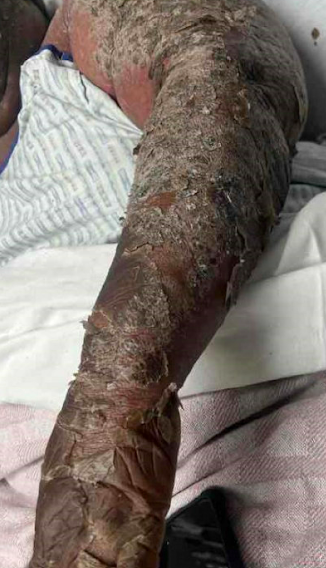
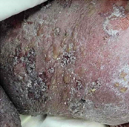
Case Presentation: A 64-year-old woman presented to the Emergency Department (ED) with widespread exanthem that started weeks prior to presentation. She reported having had similar exanthems at the same time the previous two years, which resolved with oral steroid therapy on both occasions. However, they were not as extensive as the present rash. 6 weeks before her presentation to the ED, she went to the urgent care clinic, where she received a course of methylprednisolone. She returned to the clinic with no improvement in her rash and was given a high-dose prednisone taper, which she had completed by the time of presentation to the ED. Neither a skin biopsy nor a dermatology evaluation was obtained due to social issues. On presentation, she reported that the rash became progressively more diffuse with worsening scaling and was intensely pruritic and painful. She was afebrile but was in significant distress. Skin examination showed widespread hyperpigmented patches with a hypopigmented center on a diffuse background of erythema. The lesions involved the whole body but spared the palms, soles, and genitalia. Additionally, extensive pustules and plaques were evident over the back and thighs.Ceftriaxone was commenced, given the known association of certain skin conditions with Staphylococcal superinfections.[1] A skin biopsy was obtained for pathology. The skin condition continued deteriorating, with rapidly developing flaccid blistering lesions followed by extensive desquamation involving the entire skin surface of the body and substantial exudation of serous fluid. The hospital course was complicated by shock, and she was transferred to the critical care unit (ICU) for the management of hypovolemic versus septic shock. Vancomycin was added due to concerns for Staphylococcal Scalded Skin Syndrome, provided the generalized erythematous blistering eruptions. Aggressive volume resuscitation for insensible volume losses was given. Due to concerns about hypothalamic-pituitary-adrenal axis suppression, hydrocortisone was started.The skin biopsy showed features of erythrodermic psoriasis. Treatment with cyclosporine was started, and she received wound care with triamcinolone ointment using wet wraps with sauna suit occlusion. The skin condition showed significant improvement.
Discussion: This vignette examines the case of a woman who presented with pustular psoriasis and developed erythroderma due to the receipt of systemic steroids. The appearance of the rash on the urgent care clinic visits resembled guttate psoriasis. Later, it progressively worsened, most likely due to severe flare combined with receiving steroids. The use of systemic corticosteroids in psoriasis has been reported in the literature to precipitate psoriasis flares, with the risk of developing pustular and erythrodermic psoriasis.[2] Although the likelihood of inducing a flare is low, there should always be an index of suspicion for erythroderma in deteriorating skin conditions following the administration of systemic steroids.
Conclusions: Erythrodermic psoriasis is a rare variant that involves a significant proportion of the body surface area, which is reported to be at least 75%, and requires emergency management for association with severe consequences.[3–5] The insensible fluid losses may become immense, leading to volume depletion and potentially hypovolemic shock. Another complication associated with the loss of the skin barrier is the significant risk of infection and sepsis, especially with Staphylococcal species.