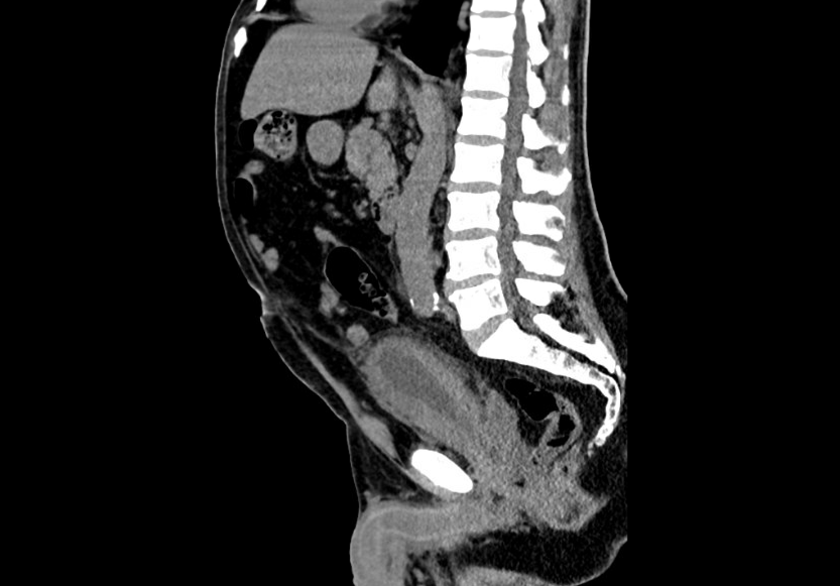
Case Presentation: A 65-year-old male with no significant past medical history was initially hospitalized for Klebsiella aerogenes urosepsis due to obstructive nephrolithiasis requiring bilateral nephrostomy tube placement and ciprofloxacin. Approximately one week later, he re-presented with severe hyponatremia. CT abdomen/pelvis revealed new thickening of the distal sigmoid colon and rectum with pelvic fat stranding. These findings were absent on imaging one month prior, which showed only bladder wall thickening. The patient had undergone unremarkable screening colonoscopy one year prior. Abdominal x-ray showed severe colonic gaseous distention, and rectal MRI revealed diffuse rectal wall edema and mesorectal enhancement. Flexible sigmoidoscopy identified a 3 cm rectal mass, with biopsy concerning for malignancy given pathologic inguinal lymph nodes. However, pathology was inconclusive due to extensive edema. Additional workup revealed bilateral lower extremity DVTs and PEs that were managed with anticoagulation. Due to extensive abdominal distention, an exploratory laparotomy was performed, revealing a 6 cm cecal serosal tear. Decompression and diverting colostomy were performed due to concern for obstructive rectal mass, and repeat biopsy was obtained. Several days later, he was readmitted with bloody nephrostomy output. CT showed new left perinephric fat stranding, progressive lymphadenopathy, and persistent bladder wall thickening. Urine culture grew Enterococcus faecium, and he was treated with linezolid. He subsequently developed an acute right-sided facial droop and weakness; MRI revealed multifocal bilateral embolic infarcts, CT abdomen identified a left renal abscess, and repeat anorectal biopsy confirmed high-grade poorly differentiated urothelial carcinoma. Due to progressive clinical decline, he transitioned to comfort focused care.
Discussion: This case highlights a rare and aggressive presentation of urothelial carcinoma with rectal metastasis, developing within months. Rectal involvement is exceptionally uncommon, with under 20 cases reported in literature (1). Due to recurrent urinary infections, bladder thickening was initially attributed to infection but likely developed due to underlying malignancy. Rapid progression to bowel obstruction, neurologic decline, and multiorgan failure underscores the need to consider malignancy in patients with hematuria, rectal bleeding, and new abdominal symptoms. While diverting colostomy is a reported palliative measure, along with chemotherapy, radiation, or resection, this patient’s hypercoagulable state led to multiple complications, precluding treatment (1-3). This case reinforces the need for consideration of aggressive malignancy and when able, rapid onset of treatment.
Conclusions: Urothelial carcinoma is the most common malignancy of the urinary tract, with bladder cancer accounting for over 90% of cases. Metastasis typically involves lymph nodes, liver, lungs, and bones; colonic involvement is rare and often indicates a poor prognosis. Metastatic disease has a 5-year 15% survival rate. (4,5) Presentation includes hematuria, urinary obstruction, or systemic symptoms. Atypical presentations can obscure diagnosis and delay treatment. This 65-year-old male’s rapidly progressive course was marked by thromboembolic events, neurologic decline, and multiorgan failure, and was ultimately found to have high-grade, poorly differentiated urothelial carcinoma with likely rectal metastasis.