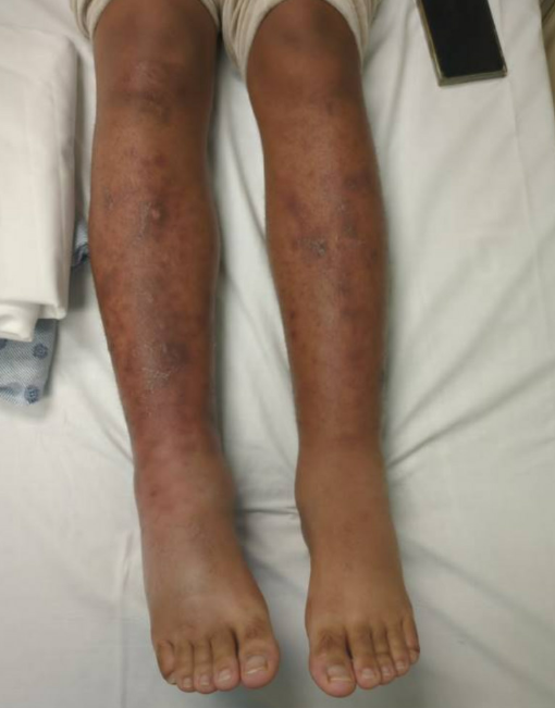
Case Presentation: A 20-year-old man with no prior medical history presented to the emergency department with acute onset of bilateral lower-extremity edema, ankle pain, purpuric lesions, erythema, and episodes of palpitations. Further history revealed subjective fevers and conjunctivitis over the preceding three days, self-administration of over-the-counter corticosteroids, and a sexual encounter with a male partner three months earlier. He denied recent travel, intravenous drug use, or animal exposure. Physical examination revealed bilateral edematous lower extremities with erythema and purpuric lesions with bilateral conjunctival injection. No significant lymphadenopathy. Laboratory evaluation revealed elevated C-reactive protein, mild anemia, a normal white blood count, creatinine within baseline, a lactate of 1.8 (mmol/L), and a urinalysis within normal limits. Bilateral lower-extremity Doppler ultrasound was negative for deep venous thrombosis. Empiric cefepime and vancomycin were initiated for presumed bilateral lower extremity cellulitis. The patient was admitted for further diagnostic workup, with considerations including cellulitis, vasculitis, Still’s disease, and bilateral conjunctivitis. During the admission, the patient’s rheumatological work-up found elevated CRP, low C4, and low cryoglobulin with negative ANA and normal C3. Infectious work-up demonstrated elevated RPR titer of 1:64, negative blood cultures, and a negative sexual transmitted infection panel. Lumbar puncture showed minimally reactive CSF Treponemal Ab-IFA, normal CSF WBC count (4 cells/µL) with 20% neutrophils, and nonreactive CSF VDRL. Skin biopsy suggested folliculitis. Ankle radiographs showed no evidence of fracture or joint effusion to suggest septic arthritis. Ophthalmology consultation revealed bilateral conjunctivitis without additional abnormalities, and a CT scan of the brain was unremarkable. Based on the patient’s ocular findings, positive RPR titer, and reactive CSF treponemal Ab-IFA, the infectious disease team made a presumed diagnosis of syphilis with concern for early neurosyphilis. The patient was discharged with penicillin G therapy and appropriate primary care follow-up.
Discussion: Syphilis is known for its varied presentations, often mimicking other conditions. In this case, atypical symptoms resembled vasculitis and cellulitis, creating diagnostic challenges. The patient’s young age and absence of hallmark features such as palmar or plantar lesions and lymphadenopathy initially suggested a rheumatologic process. However, careful review of his history and diagnostic workup—including sexual history, negative autoimmune serologies, and reactive CSF treponemal and RPR titers—was essential to establishing the diagnosis. Hospitalists should remember that while bilateral lower-extremity erythema and edema often reflect noninfectious causes such as vasculitis or stasis dermatitis, obtaining a thorough social history and keeping a broad differential for mimickers like syphilis is critical to avoid premature closure.
Conclusions: Syphilis should be considered in young adults with nonspecific systemic or dermatologic findings, particularly when initial evaluation is unrevealing. With rising incidence among young populations, hospitalists must remain vigilant in recognizing atypical presentations and interpreting serologic patterns suggestive of syphilis to ensure timely treatment.