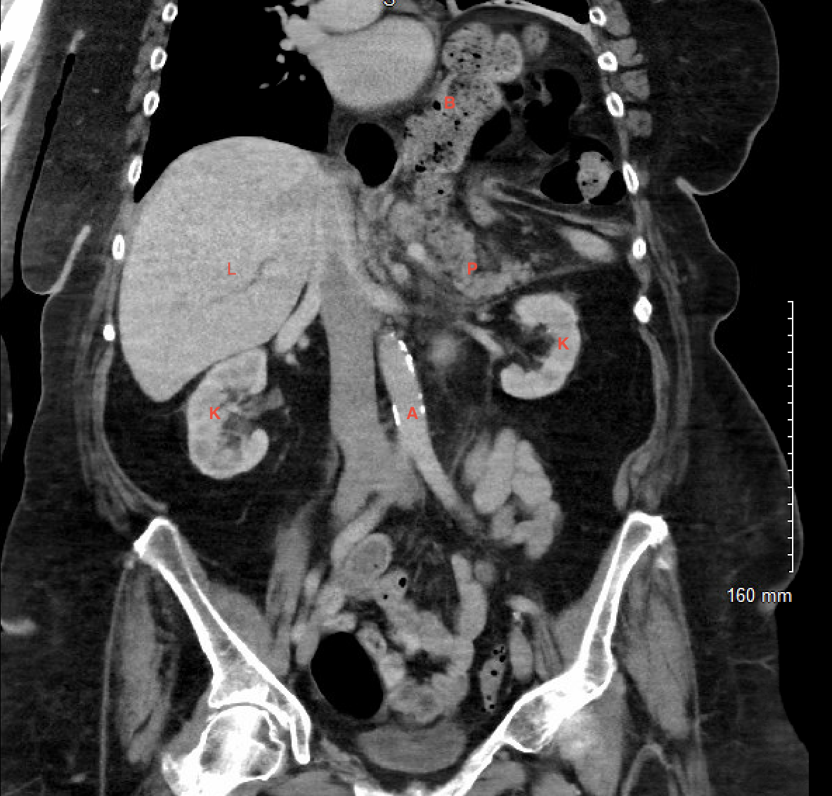
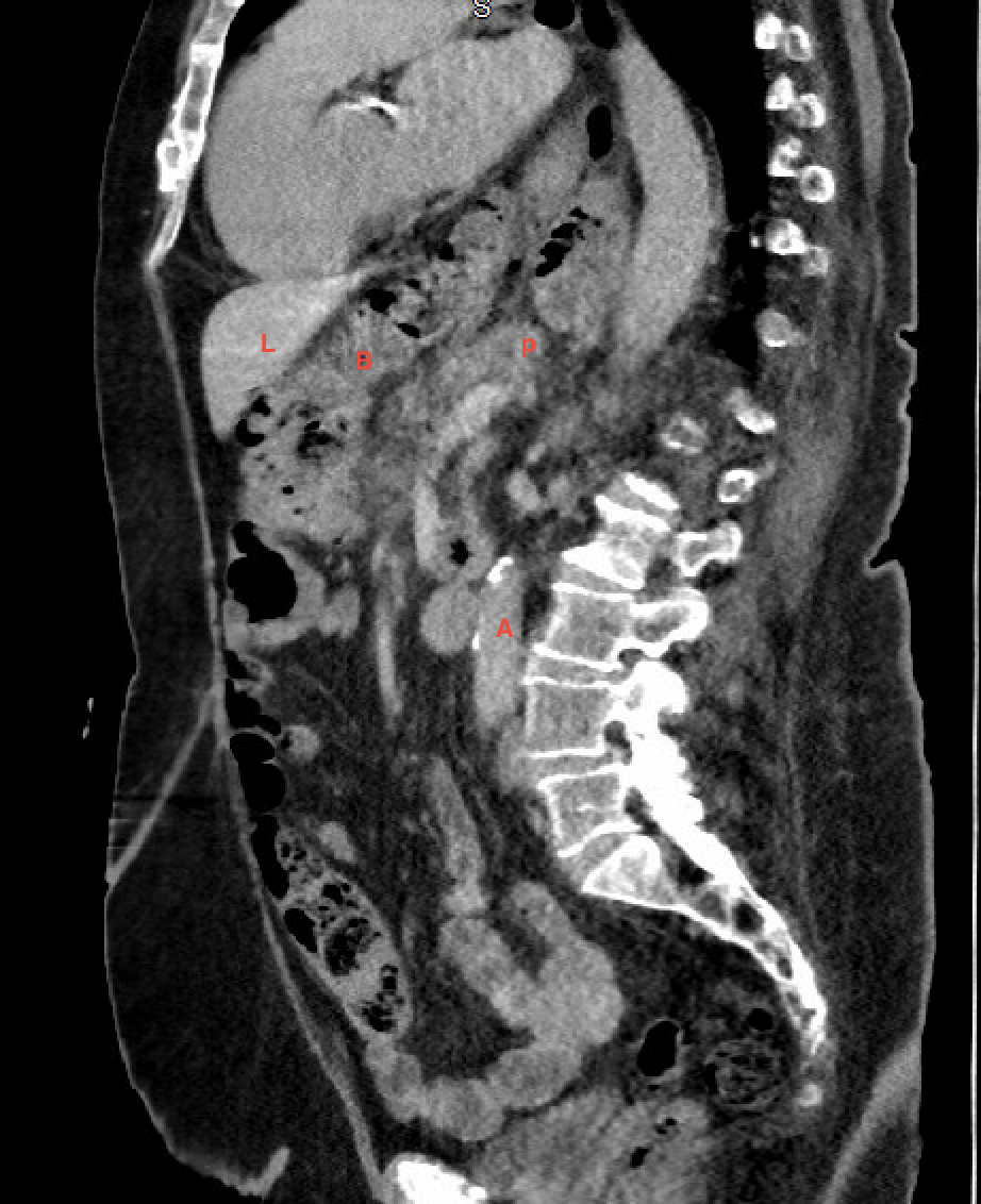
Case Presentation: A 70-year-old female with a complex medical history including Barrett’s esophagus, Class 3 obesity, obstructive sleep apnea, obesity, atrial fibrillation, and adenocarcinoma of the esophagus in remission, presented with three days of intractable nausea, vomiting, and epigastric abdominal pain. She categorized the pain as sharp and radiating to the right and left upper quadrants. Physical examination revealed epigastric tenderness. Laboratory studies showed an increased lipase level of 827 IU/L. Computed tomography (CT) scan demonstrated mild inflammatory stranding around the pancreas, attributed to low-grade pancratitis. Notably, a large paraesophageal hernia containing the stomach, bowel loops, and a slight extension of the pancreas was identified. However, there was no pancreatic necrosis or pseudocyst formation.
Discussion: Paraesophageal hiatal hernia is one of the four types of hiatal hernias, comprising 5% of all hiatal hernias. The stomach is the most common organ to herniate through the hiatal hernia, with the pancreas being the rarest due to its retroperitoneal location. This case underscores the significance of considering rare complications in paraesophageal hernias, particularly when involving uncommon organs like the pancreas. Transhiatal herniation of the pancreas can lead to disruption in the blood supply secondary to sliding through the hernia and repetitive trauma to the pancreas, resulting in acute pancreatitis. Elevation in the pancreatic enzymes, especially lipase, and radiographic evidence of inflammation and transhiatal herniation of the pancreas are diagnostic for acute pancreatitis. CT scan with IV contrast usually demonstrates the herniation and the signs of inflammation of the pancreas, as shown in the figures. While uncomplicated and asymptomatic, hiatal hernias may not necessitate intervention and are mainly managed conservatively. A high index of suspicion among physicians is crucial to diagnose and treat complications, such as acute pancreatitis and malignant transformation of the pancreas in case of a significant herniation. Treatment options for symptomatic hiatal hernias, including elective surgery, should be carefully considered, with patient preferences and risks taken into account. Continued follow-up with the surgeon is recommended for ongoing management and evaluation.
Conclusions: Pancreatic herniation through the hiatal hernia is a scarce phenomenon. Its rarity poses challenges to the diagnosis and treatment of this condition. Clinical evaluation, elevated lipase, and a CT scan of the abdomen are used to establish the diagnosis. The definitive treatment of pancreatitis secondary to paraesophageal herniation of the pancreas varies largely and depends on the patient’s comorbidities, hemodynamic stability, and preference for a surgical procedure after the resolution of pancreatitis.