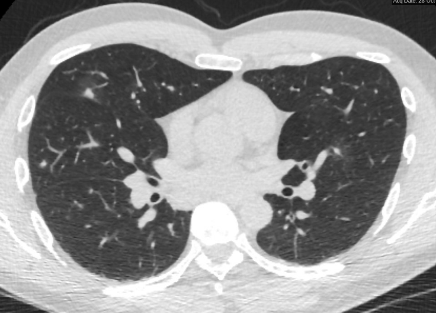
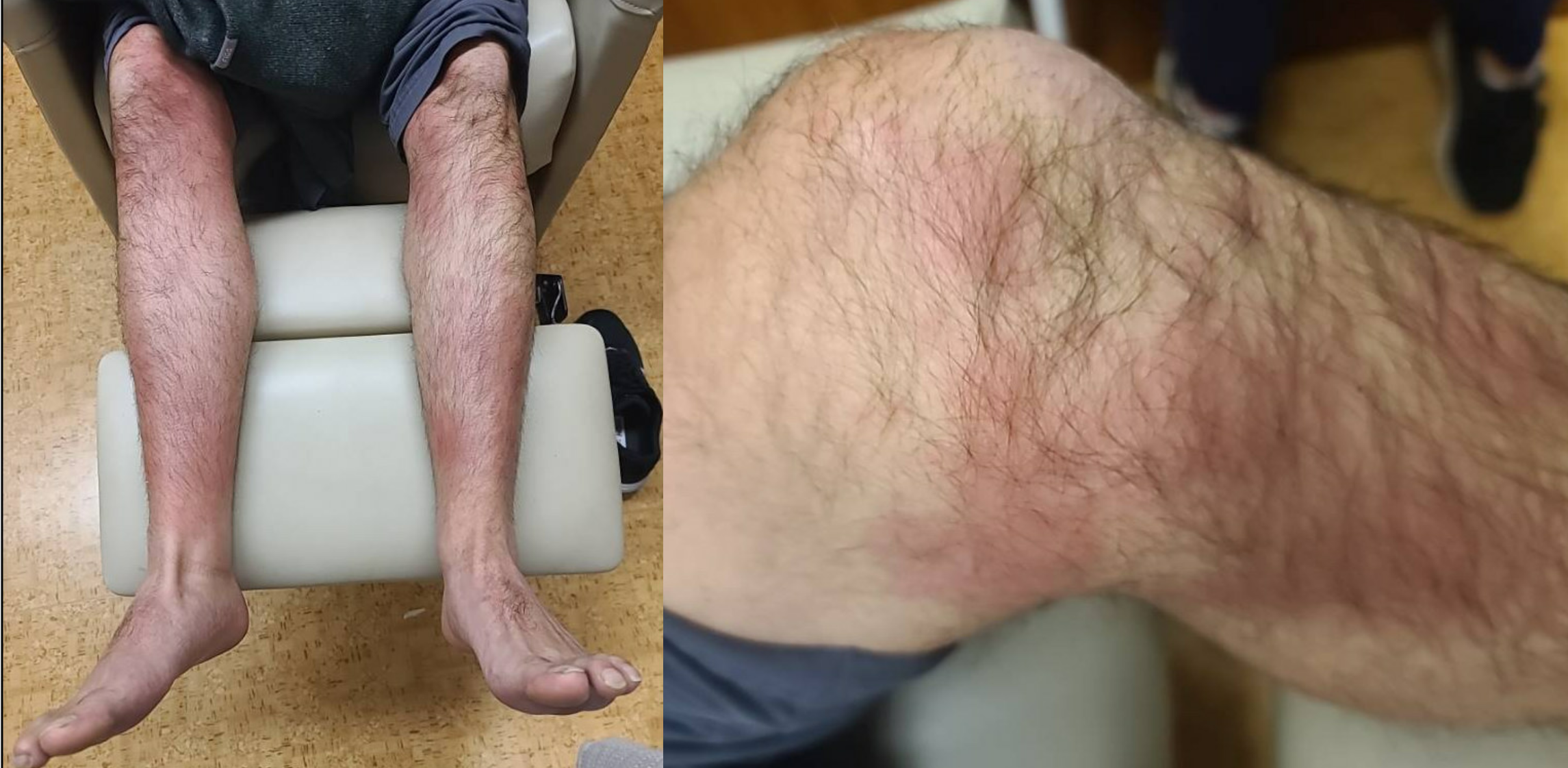
Case Presentation: A 38-year-old male dentist presented with several days of bilateral lower extremity rash and migratory large joint arthritis in the setting of fevers, myalgias, and a recent viral illness in his 3-year-old daughter. His medical history included remote testicular cancer post-orchiectomy. He was referred by his primary care provider who noted bilateral (right greater than left) knee swelling initially concerning for septic arthritis. On initial interview, he denied any recent travel, tick or insect bites, or personal history of similar rash. Initial exam was notable for temperature of 102.4°F, bilateral ankle and knee swelling, and bilateral well-demarcated, erythematous, indurated plaques most consistent with erythema nodosum. Initial chest x-ray was interpreted as normal. Punch biopsy was obtained and showed septal panniculitis and prominent eosinophils. Laboratory findings included leukocytosis (13.4) and eosinophilia, elevated C-reactive protein, and negative blood cultures. Fevers persisted throughout hospitalization despite empiric treatment with doxycycline and ceftriaxone. Given unclear etiology, additional history was collected with the patient’s wife present who endorsed recent work-related travel to Arizona that the patient did not recall in the setting of his acute illness, despite otherwise full orientation. On further questioning, he reported that his hotel was near an active construction site and he frequently kept the window open. With new elements to his history, CT chest was obtained and demonstrated numerous pulmonary nodules with peripheral groundglass halos. Serum Coccidioides antibody was found to be positive. He was initiated on fluconazole with interval improvement in symptoms.
Discussion: Here we present a case of coccidioidomycosis characterized by rare immunologic manifestations including erythema nodosum and symmetrical arthralgias, as well as diffuse reticulonodular pneumonia in an immunocompetent adult in a non-endemic location. We postulate his severe symptoms were a consequence of high inoculum dose in a host without previous exposure, which can worsen symptoms in certain other conditions such as malaria. Initial exposure history obtained by multiple providers did not elucidate an explanation for this patient’s protean symptoms. Continued patient interview and family collateral revealed an exposure event due to disturbed soil in the American southwest. This highlights the importance of an accurate initial history and the need to collect more information, including collateral from family, when the etiology of persistent fever is unclear. This new evidence prompted reconsideration of his chest x-ray, on which multiple nodular opacities were now appreciated.
Conclusions: Undifferentiated, persistent fever requires accurate, and often repeated, history taking, and this patient with endemic coccidioidomycosis was only diagnosed after multiple interviews and family collateral.