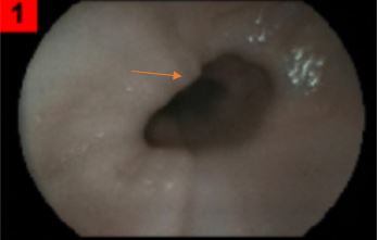
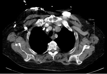
Case Presentation: A 57-year-old incarcerated male with end stage renal disease (ESRD) on hemodialysis, coronary artery disease (CAD), and Gastroesophageal reflux disease (GERD) presented to the emergency department with several episodes of hematemesis, shortness of breath and fatigue for the past 2 days without any associated abdominal pain, dysphagia, or odynophagia. On exam, he was afebrile and hypertensive without tachycardia. He had dry mucous membranes and abdominal and skin exam did not reveal any stigmata of chronic liver disease. Laboratory evaluation was significant for macrocytic anemia (hemoglobin 9.7 gm/dL), elevated BUN (75 mg/dL), elevated creatinine (8.71 mg/dL), anion gap metabolic acidosis (24) with an elevated lactic acid (6.2 mmol/L). After fluid resuscitation, EGD was performed the following day which revealed Grade II varices in the upper 1/3 of the esophagus (Figure 1) and he underwent subsequent variceal band ligation. Given his history of chronic hemodialysis with repeated central line placements, superior vena cava (SVC) obstruction was suspected. CT scan of the chest, abdomen, and pelvis with oral and IV contrast revealed stenosis of the SVC and right internal jugular vein (IJV) with dilated chest wall veins (Figure 2). Patient remained stable and was discharged with the plan for SVC stenting and/or angioplasty on an elective basis.
Discussion: “Downhill” or upper esophageal varices (EV) are believed to comprise about 0.1% of all esophageal varices [1,2]. EV are most commonly located in the distal esophagus and the majority of which result from portal hypertension. Less commonly, upper EV can be present in the upper esophagus or may involve the entire esophagus. Their presence does not denote cirrhosis or portal hypertension like distal EV. When a diagnosis of a downhill varix is made, it should prompt evaluation for venous obstruction in the vessels that drain the esophagus. These vessels are numerous and are as follows: The upper esophageal plexus drains into the inferior thyroid veins. The middle esophageal plexus drains into the azygos, hemiazygos, bronchial, and intercostal veins. All the previous veins drain into the SVC. An obstruction in any of the aforementioned veins can cause upper esophageal varices. SVC angioplasty with stent placement has been effective at reducing formation of varices in patients with SVC stenosis [4,5].The most common cause of downhill varices is a result of repeated central venous catheter placement with resultant SVC stenosis [6]. Other important differential diagnoses that should be considered are lung, thymic, or thyroid malignancies. One should also consider mediastinal fibrosis in patient’s with prior history of radiation or castleman’s disease in patienta with enlarging lymph nodes and B symptoms.
Conclusions: Upper esophageal varices are an extremely rare entity, and although they only represent 0.1% of all varices, distinguishing between an upper and lower varix is vital. It not only will guide treatment, but the two are unique entities which have their distinct etiologies. Definitive treatment includes identifying the underlying etiology. For surgical candidates, stenting or angioplasty is the mainstay of treatment, while nonsurgical candidates can benefit from variceal band ligation or sclerotherapy as a temporizing measure.