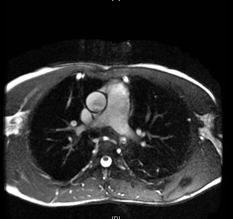
Case Presentation: An 18 year old Spanish speaking male from Honduras presented following a motor vehicle accident. Initial assessment in the ED included a CXR and bedside Echocardiogram which noted cardiomegaly and a reduced EF of 15%. Patient reports experiencing headaches, blurry vision, palpitations, and non-radiating left sided chest pressure intermittently over the last four years. He reported never having seen a physician and denied any family history of cardiac disease or sudden cardiac death. Physical was exam notable for systolic hypertension in the upper extremities, systolic ejection murmur left sternal border, diminished femoral pulses, and thin lower extremities. The patient reported no other significant past medical history. 24-hour urine fractionated metanephrines and catecholamines were within normal limits, HIV and T. Cruzi antibodies were negative. Transesophageal Echo and Cardiac MRI revealed a bicuspid aortic valve with fusion of the right and left coronary cusps, moderate-severe eccentric aortic regurgitation, severe global LV systolic dysfunction, and coarctation of the aorta. He subsequently underwent AV repair, Descending Aorto-bypass graft, and Coarctation of Aorta repair. Follow up Echo revealed elevated mean transaortic valve gradient in the presence of a bioprosthetic aortic valve and severely dilated left atrium.
Discussion: Coarctation of the aorta is a discrete aortic narrowing, located beyond the left subclavian artery, causing hypertension proximal and reduced blood pressure distal to the aortic narrowing. Patients with aortic coarctation may be asymptomatic or present with hypertension, symptoms of exertional leg fatigue, or headaches. The mechanism for hypertension is a combination of the mechanical obstruction itself, the high output into the accessory collateral circulation, and the renin-angiotensin-aldosterone system. Upper extremity hypertension and reduced blood pressure and pulse amplitude in the lower extremities are characteristic findings which in turn, cause a radial artery to femoral artery pulse delay. The patient was not found to have any systemic disorder, such as connective tissue disease, rheumatic or infectious aortitis leading to the presumption that this was a congenital disorder.
Conclusions: The delayed presentation was of particular interest to the treating team as it hallmarked the paradox of health care inequality. Use of advanced cardiac imaging captured a diagnosis that was likely missed due to lack of contact with the health care system in his native country. As demonstrated in this case, there is no one-size-fits-all differential when it comes to considering a new diagnosis of dilated cardiomyopathy in a young patient. It is incumbent upon clinicians to suspend skepticism and entertain a broader differential when caring for patients with limited access to proper medical care. One such example, is a mechanical coarctation of the aorta as the etiology for heart failure in a young adult. Clinicians must remain attentive caring for any patient disconnected from the health care system, as they may present with a seemingly improbable diagnosis, only identified by clinicians keen enough to look for it. The case is also a call for academic physicians to find novel ways to care for those with limited access to health care, both locally and abroad.