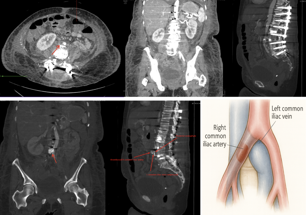
Case Presentation: A 73-year-old woman with Charcot-Marie-Tooth disease underwent T11-pelvis spinal fusion in October, complicated by a surgical site infection. In late December, she was diagnosed with a left peroneal vein deep vein thrombosis (DVT) and started on apixaban. One week later, she returned to the emergency department with worsening left lower extremity swelling and erythema, concerning for cellulitis (Fig. 1). Despite broad-spectrum antibiotics, her symptoms persisted, prompting further imaging. Venous duplex demonstrated a persistent distal peroneal DVT, and CT abdomen/pelvis revealed non-visualization of the right common and external iliac arteries concerning for thrombosis, which was subsequently confirmed on CT angiogram (Fig. 2).Interventional radiology was consulted and identified compression of the left common iliac vein (LCIV) between the thrombosed right common iliac artery (RCIA) anteriorly and a postoperative osteophyte complex posteriorly. May-Thurner Syndrome (MTS) was suspected, though there was no evidence of more proximal thrombosis in the iliofemoral venous system. The patient’s symptoms resolved with continued antibiotics and anticoagulation, and a repeat venous duplex demonstrated resolution of the distal DVT. She was later referred for outpatient venography to evaluate the need for iliac venous stenting, which she declined, preferring to avoid further invasive interventions. She remains clinically stable and is managed conservatively with physical therapy.
Discussion: MTS, or iliac vein compression syndrome, occurs when the LCIV is narrowed by overlying structures, most commonly by the RCIA. Although often underrecognized, MTS can be induced or exacerbated by spinal pathology, such as osteophytes, or by prior lumbar surgery. Postoperative hardware, retroperitoneal scarring, bony changes, and iatrogenic alterations have all been reported as additional compressive forces that further compromise venous outflow and hemodynamics [2–6]. In most cases, venous thrombosis rather than arterial thrombosis is implicated in MTS. To our knowledge, this is the first reported case of RCIA thrombosis, in combination with postoperative bony changes, producing dual compression of the LCIV. This anatomical abnormality and atypical mechanism explain the patient’s persistent unilateral swelling and lack of improvement despite initial intervention.Recognition of MTS is clinically important, as untreated cases may result in chronic venous insufficiency, recurrent thrombosis, or pulmonary embolism. This case illustrates how postoperative anatomic changes can intensify or unmask underlying venous compression, creating a complex clinical picture that requires further investigation.
Conclusions: Although MTS is estimated to account for 2% to 5% of all DVTs [1], its true prevalence is likely much higher and remains underrecognized in current literature [2]. For this reason, this venous anatomical variation should be considered in patients with unilateral lower extremity swelling following lumbar spine surgery, particularly when symptoms persist despite standard treatments. Postoperative anatomic changes may intensify or reveal underlying iliac vein compression, predisposing to MTS. Early recognition and timely endovascular evaluation can prevent recurrent thromboembolism, reduce diagnostic delays and rehospitalizations, and mitigate long-term morbidity, making MTS an especially relevant entity for hospital medicine.
.png)