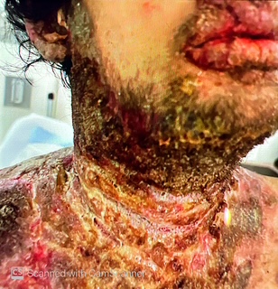
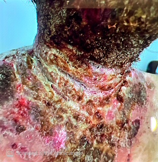
Case Presentation: Case PresentationA 27-year-old man with eczema and polysubstance use disorder presented with a two-month history of a progressively worsening rash. The eruption began as pruritic raised lesions on the scalp while living in Connecticut and gradually extended to the peri-auricular region, neck, chest, abdomen, and right axilla. A brief course of oral antibiotics from an urgent care visit led to transient improvement, but symptoms recurred and rapidly progressed after discontinuation. He reported no recent travel, sick contacts, or new medications and had not sought dermatologic care due to lack of insurance.On arrival, he was tachycardic to the 120s; toxicology was positive for cocaine. Dermatology and infectious disease consultations were obtained, and empiric intravenous vancomycin and cefepime were started for presumed severe skin and soft-tissue infection. Laboratory evaluation showed leukocytosis, anemia, hypoalbuminemia, hyponatremia, and hypocalcemia. He met sepsis criteria on the basis of tachycardia and leukocytosis. A punch biopsy was performed.Dermatopathology demonstrated features consistent with pemphigus vegetans, including epidermal acanthosis, suprabasal acantholysis, and eosinophilic microabscesses. Bacterial cultures from lesional skin grew methicillin-sensitive Staphylococcus aureus and Pseudomonas aeruginosa, indicating polymicrobial superinfection.Systemic prednisone and high-potency topical corticosteroids were initiated. Vancomycin was discontinued after two days; cefepime was continued for seven days before transition to oral cephalexin and ciprofloxacin. Nutritional assessment identified mild protein-calorie malnutrition, and supplementation was begun. Because of the known association between pemphigus vegetans and inflammatory bowel disease, gastroenterology follow-up was arranged. Addiction-medicine referral was provided at the patient’s request.Over the hospitalization, the rash improved markedly with corticosteroids, and his tachycardia resolved. He was discharged on a prednisone taper, topical corticosteroids, and oral antibiotics, with outpatient follow-up across dermatology, infectious disease, gastroenterology, and addiction medicine.
Discussion: DiscussionPemphigus vegetans is a rare variant of pemphigus vulgaris and often presents with vegetating plaques in intertriginous regions. Delayed diagnosis is common when lesions mimic infectious or eczematous dermatitis. Superimposed bacterial infection, particularly with S. aureus or Pseudomonas, can intensify inflammation and contribute to systemic findings, as in this case. Systemic glucocorticoids are first-line therapy, with biopsy essential for diagnostic confirmation. Screening for associated autoimmune conditions is recommended.
Conclusions: ConclusionThis case illustrates a rapidly progressive presentation of pemphigus vegetans complicated by polymicrobial superinfection, initially resembling a severe infection. Early biopsy, targeted antimicrobial therapy, and timely immunosuppression were key to clinical improvement and highlight the importance of multidisciplinary care and addressing barriers to access in complex dermatologic disease.