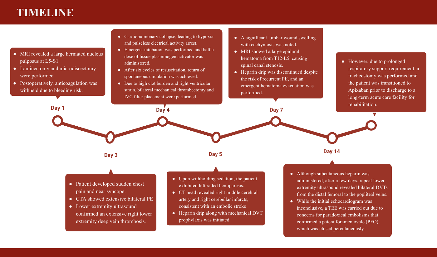
Case Presentation: Pulmonary embolism (PE) is a major cause of sudden cardiac arrest and circulatory collapse in patients under the age of sixty-five. We present a case of a young adult with recurrent venous thromboembolisms (VTE) who developed PE and stroke due to the inability to continue anticoagulation postoperatively. A 37-year-old male with recurrent VTE on anticoagulants and family history of hypercoagulability presented with worsening back, numbness and bowel and bladder incontinence for a month. Magnetic resonance imaging (MRI) revealed a large herniated nucleus pulposus at L5-S1, and laminectomy and microdiscectomy were performed. Postoperatively, anticoagulation was withheld due to bleeding risk. Within 48 hours, the patient developed sudden chest pain and near syncope. Computed tomography (CT) angiography showed extensive bilateral PE, and a lower extremity ultrasound confirmed an extensive right lower extremity deep vein thrombosis (DVT). He experienced cardiopulmonary collapse, leading to hypoxia and pulseless electrical activity arrest. Emergent intubation was performed and half dose of tissue plasminogen activator was administered. After six cycles of resuscitation, return of spontaneous circulation was achieved. Due to high clot burden and right ventricular strain, bilateral mechanical thrombectomy and IVC filter placement were performed. Upon withholding sedation, the patient exhibited left-sided hemiparesis. CT head revealed right middle cerebral artery and right cerebellar infarcts, consistent with an embolic stroke, and a heparin drip along with mechanical DVT prophylaxis was initiated. Shortly after, a significant lumbar wound swelling with ecchymosis was noted. MRI showed a large epidural hematoma from T12-L5, causing spinal canal stenosis. Heparin drip was discontinued despite the risk of recurrent PE, and an emergent hematoma evacuation was performed. Although subcutaneous heparin was administered, after a few days, repeat lower extremity ultrasound revealed bilateral DVTs from the distal femoral to the popliteal veins. While the initial echocardiogram was inconclusive, a transesophageal echocardiogram was carried out due to concerns for paradoxical embolisms that confirmed a patent foramen ovale (PFO), which was closed percutaneously. Due to prolonged respiratory support requirement, a tracheostomy was performed and the patient was transitioned to Apixaban prior to discharge to a long-term acute care facility for rehabilitation.
Discussion: Due to the high fatality risk and complications associated with PE, early identification and prompt management are crucial. However, in this case, the absolute contraindication of recent spinal surgery necessitated withholding anticoagulation, delaying timely treatment. Additionally, the patient’s susceptibility to recurrent VTEs contributed to ongoing complications. Literature suggests an increased risk of stroke in patients with acute PE, likely due to paradoxical embolism via a PFO. A multidisciplinary approach, including a PE response team (PERT), facilitates rapid intervention and protocol-driven care. However, given the diverse presentations and potential complications, treatment must be tailored to each individual patient.
Conclusions: This case highlights the challenges of managing VTE in patients with contraindications to anticoagulation, the need for a multidisciplinary approach and an individualized, protocol-driven management to balance thrombotic and bleeding risks effectively.