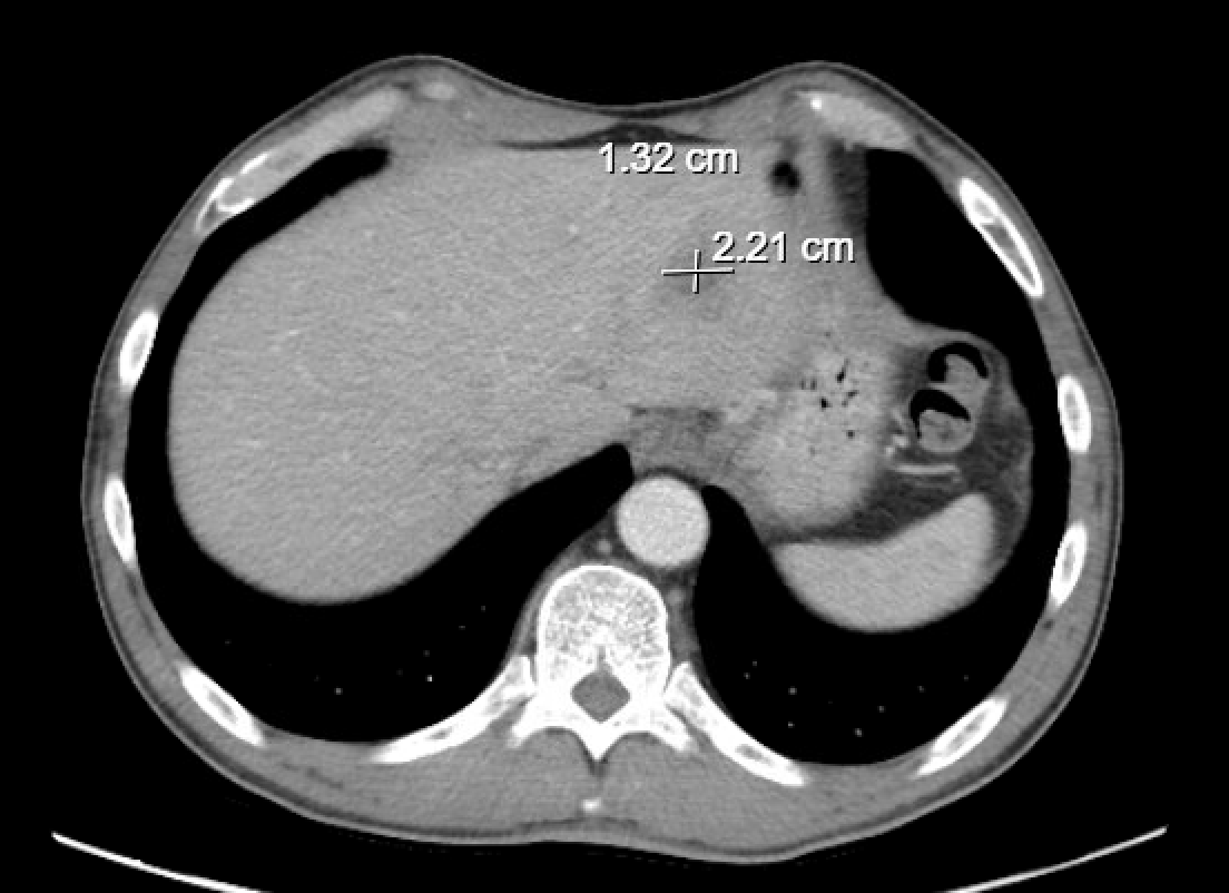
Case Presentation: A 57-year-old man with a history of an intra-abdominal abscess (2012), appendectomy, and prior orthopedic surgeries presented with 8 days of profound fatigue, fevers (Tmax 104°F), malaise, and progressive left-sided abdominal pain. He is a triathlete with baseline physiologic bradycardia but was noted to have a persistently higher heart rate (80–90s) and dyspnea on exertion. He denied nausea, vomiting, diarrhea, or abdominal distension. He reported no recent travel aside from trips to Mexico approximately one year prior and Costa Rica six months prior, during which he remained asymptomatic.On admission, he was febrile and uncomfortable but hemodynamically stable. Laboratory evaluation revealed leukocytosis and elevated inflammatory markers. CT imaging demonstrated a hepatic abscess, and MRI confirmed a loculated hepatic lesion. Interventional radiology performed percutaneous drainage of purulent material. Despite two attempts, abscess fluid cultures remained negative, as did blood cultures. Given the persistently sterile cultures, Infectious Disease consultants recommended gastrointestinal PCR testing of the drainage fluid, which was positive for Entamoeba histolytica. Subsequent testing demonstrated positive serum IgG and stool PCR for E. histolytica. A later culture from the drainage fluid yielded P. curis.The patient received IV piperacillin–tazobactam for nine days, after which he was transitioned to oral levofloxacin and metronidazole. A seven-day course of paromomycin was prescribed for luminal eradication. The hepatic drain was removed before discharge once output had ceased.
Discussion: The patient remained hospitalized for 12 days because the causative organism of his hepatic abscess was initially unidentified. The delay in diagnosing amebic liver abscess led to unnecessary prolonged IV antibiotic therapy and extended hospitalization despite the patient otherwise feeling clinically well.This case underscores three key diagnostic pitfalls. First, among travelers and migrants, only a minority of individuals with Entamoeba histolytica infection develop gastrointestinal symptoms, contributing to a low index of suspicion in non-endemic regions. Second, a negative abscess fluid culture does not exclude amebiasis; hepatic abscess contents are typically sterile, consisting predominantly of necrotic hepatocytes and debris, with few inflammatory cells and only peripheral trophozoites. Serology or serum antigen detection is the most informative noninvasive diagnostic tool for distinguishing E. histolytica liver abscess from pyogenic etiologies. In the United States and other low-prevalence settings, multiplex GI PCR testing of stool or abscess fluid can rapidly and accurately identify E. histolytica with high predictive value due to low background prevalence. Third, delayed onset after remote travel is common. Although most cases present within 2–5 months of exposure, amebic liver abscess has been reported months to years after travel to endemic areas, further complicating recognition.
Conclusions: Patients presenting with a hepatic abscess and a history of remote travel should be evaluated for amebic liver abscess even in the absence of diarrhea. This case underscores the importance of obtaining a comprehensive travel history and highlights the diagnostic utility of early serologic testing and GI PCR in non-endemic regions, where rapid identification of E. histolytica can prevent delays in targeted therapy.
.png)