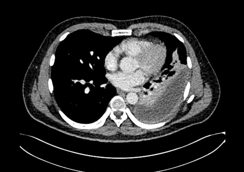
Case Presentation: A 52-year-old male with history of hypertension, GERD, and recently treated outpatient pneumonia presented with fevers (103F), progressive dyspnea on exertion, left-sided chest pain, and night sweats. Three weeks ago, he first noted reproducible left-sided chest pain and was evaluated in the emergency department with a normal ECG. Two weeks prior, he re-presented with ongoing left-sided chest pain and fever. CTA showed multifocal lower lobe pneumonia and a small left-sided effusion. He was prescribed azithromycin and cefdinir then prednisone for ongoing symptoms. He denied diarrhea, rash, recent travel, dental infections, and substance use. On vitals, he was tachycardic (102 bpm), normotensive (135/75 mmHg), and febrile (38.8C). Exam was notable for tachypnea and diminished left breath sounds. On labs, he had leukocytosis (WBC 19.4 10*9L), thrombocytosis (495 10*9/L), hypoalbuminemia (2.9g/dL), and elevated liver enzymes (ALT 109 IU/L, alk phos 378 IU/L). Respiratory pathogen panel and COVID test were negative. CT revealed a loculated left parapneumonic effusion and an enlarged hepatic lesion concerning for hemangioma. Broad-spectrum antibiotics (vancomycin, cefepime, and metronidazole) were started for sepsis concern. Interventional pulmonology was consulted and placed a chest tube, draining 400ml of purulent pleural fluid. Pleural studies revealed an exudative effusion, and cultures grew Fusobacterium nucleatum and Streptococcus anginosus confirming an empyema. Blood cultures were negative. He received six doses of intrapleural fibrinolytic during admission. On Day 5, with reduced fluid output, serosanguinous drainage, and marked radiologic improvement, he was transitioned to oral amoxicillin/clavulanate for 6-week course. Outpatient chest X-ray demonstrated left lower lobe volume loss with scarring but no new consolidation.
Discussion: In this case, diagnostic delay led to development of an empyema. First, the patient had a delayed diagnosis of pneumonia then had findings of a small effusion on CT scan but further evaluation and treatment were not pursued. In all cases of pneumonia with effusion, chest ultrasonography should be performed to evaluate size and signs of loculations. If able, the patient should receive a thoracentesis to evaluate for complex effusion 1. Although anaerobic coverage is not recommended for uncomplicated community acquired pneumonia, it should be given in cases where a complex effusion of empyema is suspected 2.Fusobacterium nucleatum is an anaerobic Gram-negative bacilli commonly in the oral cavity. Though typically commensal, it can act as an opportunistic pathogen and has been implicated in a range of human diseases such as periodontitis, adverse pregnancy outcomes, and Lemierre’s syndrome. It is one of the most frequently identified anaerobic species in empyema 3. Clinical presentation is often nonspecific and can mimic typical bacterial pneumonia, leading to delays in diagnosis and targeted therapy. However, once identified it can be effectively treated with first-line antibiotic agents: metronidazole, β-lactam/β-lactamase inhibitors, and carbapenems4.
Conclusions: This case highlights the importance of considering empyema and anaerobic pathogens such as F. nucleatum in patients with a prolonged pneumonia and clinical worsening. A thorough workup with risk factor assessment, appropriate antibiotic coverage, and timely drainage are critical for favorable outcomes in anaerobic empyema.