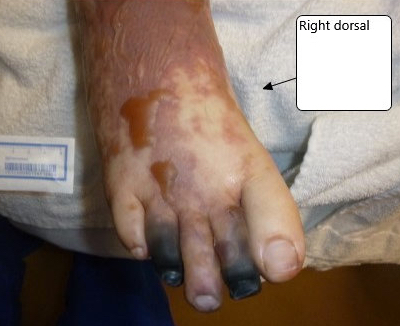
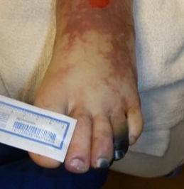
Case Presentation: A 39-year-old male post-splenectomy due to hereditary spherocytosis presented with syncope, fatigue, fever and dark urine. He had associated tachycardia and fever. Physical exam was unremarkable. Labs showed an elevated lactate, creatinine, bilirubin, transaminases, and markedly elevated D-dimer. CBC was normal. He received IV fluid resuscitation and IV Rocephin. The patient subsequently desaturated and received empiric IV heparin for suspected pulmonary embolism. PT, PTT, and INR were prolonged. Lactic acid continued to rise, and renal function deteriorated. CRP was elevated but ESR was normal. Blood cultures grew Streptococcus pneumoniae. He developed mottling throughout the extremities with associated pain. The patient then developed severe thrombocytopenia and low fibrinogen. He received fresh frozen plasma and protein C (PC) concentrate. Hemodialysis was initiated for anuria and he suffered multiple necrotic digits from worsening purpura fulminans (PF). Blood smear revealed schistocytes. Antithrombin III and PC activity were low. On day three, his D-dimer continued to rise. LDH was elevated with a normal reticulocyte count. Hematology recommended urgent transfer to our facility for emergent therapeutic plasma exchange (TPE). Upon transfer, patient improved markedly after initial TPE. ADAMTS13 activity ruled out thrombotic thrombocytopenic purpura (TTP). Sepsis-related disseminated intravascular coagulation (DIC) was diagnosed. He received five TPE cycles prior to full recovery.
Discussion: This case highlights the diagnostic challenge in distinguishing between thrombotic microangiopathies (TMAs) and DIC. Although initial TTP suspicion was ruled out, the patient’s TPE response implies a broader diagnosis beyond sepsis-related DIC with PF. Prompt recognition and targeted interventions, including TPE, are essential in such cases. Differential diagnosis of TMAs, particularly TTP, remains crucial in cases of severe thrombocytopenia and hemolytic anemia. The PLASMIC score aids in early identification, and TPE stands as a pivotal intervention, removing autoantibodies and large von Willebrand factor multimers. In the context of DIC, heparin may be considered, and studies suggest TPE’s efficacy in sepsis-associated DIC, possibly through the removal of inflammatory cytokines and restoration of endothelial function. PF, associated with DIC, is a life-threatening syndrome, and our case’s occurrence following Streptococcus pneumoniae infection aligns with known infectious etiologies. TPE was chosen in our case as an urgent intervention due to the patient’s rapidly deteriorating condition, clinical suspicion of TMA, and the need for quick removal of potential pathogenic factors. Timely TPE was pivotal in reversing clinical decline and underscores its significance. Studies affirm TPE’s efficacy in TTP and sepsis-associated DIC.
Conclusions: In critically ill patients, distinguishing between TMA and DIC poses diagnostic and therapeutic challenges. Timely recognition and targeted interventions, particularly the use of TPE, can be pivotal in reversing patient’s rapid clinical decline. Complex coagulopathy cases require a timely and comprehensive approach.