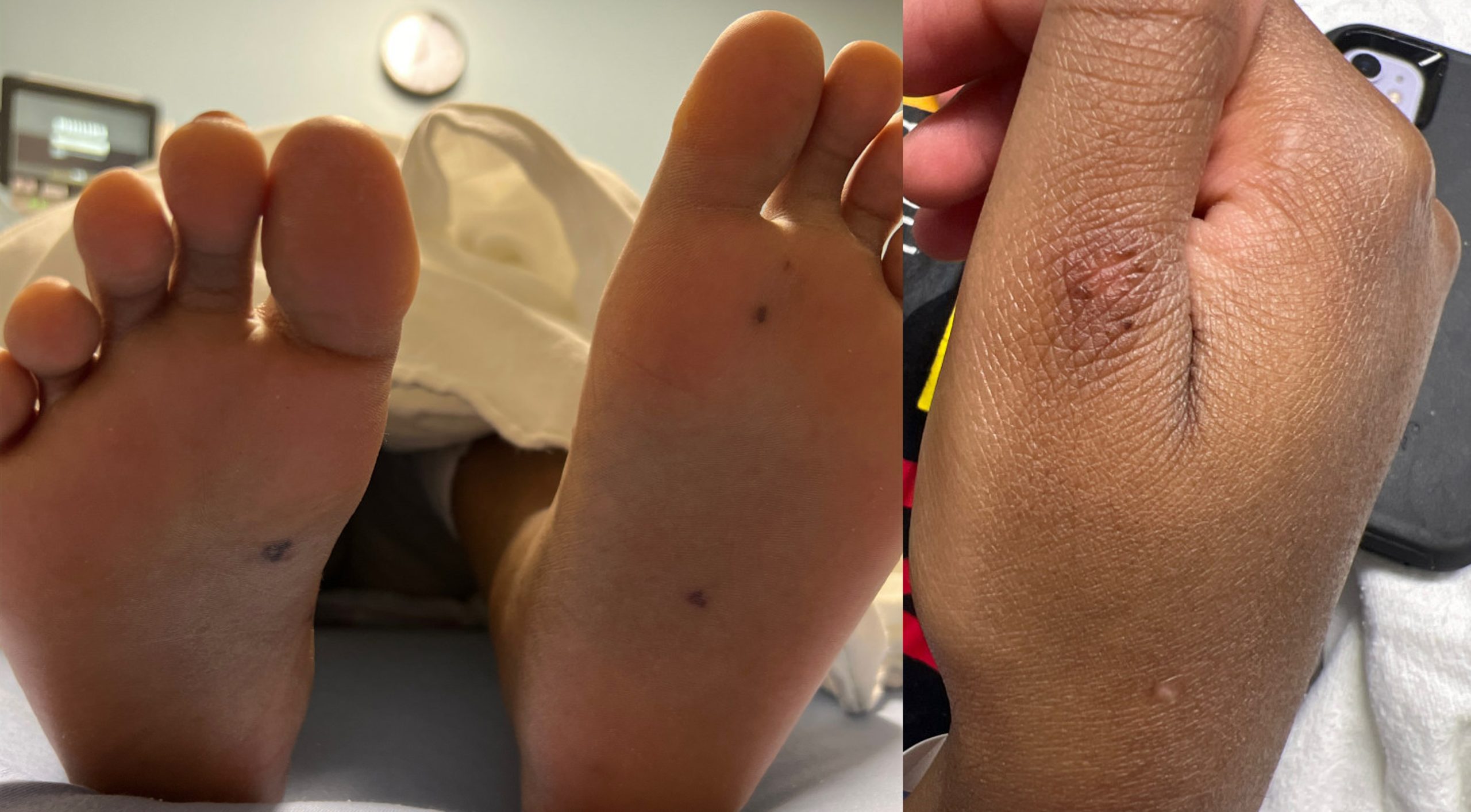
Case Presentation: A 17 y.o. female with systemic lupus erythematosus (SLE) presented with several weeks of worsening migratory polyarthralgia, as well as sporadic fevers, night sweats, and chills. Prednisone was begun 3 weeks prior for a presumed SLE flare, given elevated anti double-stranded DNA (anti-dsDNA) and erythrocyte sedimentation rate (ESR) and hematuria, but provided no relief. Prior flares featured malar rash, poor appetite, and less predominantly joint pain. She reported last sexual activity a year prior, with one lifetime partner and use of condoms. She was afebrile on presentation. Exam found no arthritis but was notable for small, hemorrhagic lesions of her feet, ankles, and right thumb (Figure). Labs found leukocytosis with neutrophilia and elevated CRP, atypical of her SLE flares. Anti-dsDNA and ESR were elevated but improved. Echocardiogram was normal without vegetations. Nucleic acid amplification testing (NAAT) was positive for gonorrhea on throat swab but negative in urine. She was treated with 7 days of ceftriaxone with rapid symptom resolution. Repeat gonorrheal NAAT of throat and urine were negative, blood cultures were negative, and her partner sought treatment independently.
Discussion: This case describes a disseminated gonococcal infection (DGI) masquerading as an SLE flare. Hospitalists should remain vigilant for atypical or fulminant presentations of infectious disease in SLE patients, given their complement depletion and immunosuppression, and should keep a broad differential for arthralgia if findings are inconsistent with prior flares. DGI occurs historically in 0.5-3.0% of patients with Neisseria gonorrhoeae infection, in 0.6-1.7% of patients with septic arthritis, and classically in women younger than 40 y.o. but with increasing incidence in older patients and men. Phenotypes include an arthritis-dermatitis syndrome (with polyarthralgia, dermatitis, and tenosynovitis) and a more isolated purulent arthritis. Dermatitis occurs in 75% of patients, with lesions primarily in distal extremities, typically numbering 2-10, often transient, and with varying morphisms (classically painless and pustular, but also hemorrhagic macules, papules, bullae, or nodules). Cultures of disseminated sites are often negative. Gold standard for diagnosis is NAAT of urine or vaginal swab (and urogenital, rectal, and pharyngeal sites as indicated), but clinical diagnosis can be made with rapid response to third-generation cephalosporins. Rare complications include endocarditis, meningitis, osteomyelitis, and abscesses. Of note, this patient initially reported no current sexual activity but had positive NAAT on throat swab and a current oral sex partner, highlighting the importance of collecting a thorough sexual history.
Conclusions: Hospitalists should maintain a broad differential for arthralgias, be aware of typical skin findings for many diseases including DGI, and remain especially vigilant with patients with SLE, a disease known for protean manifestations. In immunosuppressed SLE patients with findings inconsistent with prior flares, high suspicion should remain for alternate diagnoses, particularly for advanced or disseminated infectious diseases. A thorough and specific sexual history is essential to developing a complete differential diagnosis. DGI presentations are variable with transient features, but rapid improvement with third-generation cephalosporins can be clinically diagnostic.