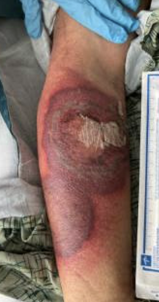
Case Presentation: A 45 year old male with relapsed refractory AML status post multiple lines of therapy including stem cell transplant (most recently on revumenib), with concern for graft versus host disease of the skin (on ruxolitinib and prednisone), was admitted for neutropenic fever. His BP was 104/68 and respiratory rate was 20. On exam he had left axillary lymphadenopathy and a ~7 cm sized erythematous patch on his right lower extremity, thought to be cellulitis (figure 1). Otherwise, exam and vitals were normal. The patient was profoundly pancytopenic: ANC 200 cells/mm3, hemoglobin 7.9 g/dL, and platelets 10,000 cells/mm3. Blood cultures were negative throughout admission. Broad spectrum antibiotics with isavuconazonium, cefepime, and vancomycin were started and prophylaxis of atovaquone and acyclovir was continued. His cellulitis initially improved, but on day three he developed central darkening and lesion growth (figure 1). He also developed several new pruritic 1-2 cm, dark, erythematous macules on his face and head, and in later days developed similar lesions on his neck, shoulders, and limbs. These lesions grew, and had some desquamation (figure 2). He was started on amphotericin B and micafungin for concern for angioinvasive infection. Dermatology biopsied a lesion, which showed mixed inflammatory cell infiltrates with many neutrophils, lymphocytes, and histiocytes. While indeterminant, the lack of biopsy culture growth was more consistent with Sweet Syndrome (SS). Karius testing was sent to rule out angioinvasive infection and was negative, as were fungitell and galactomannan, so antifungals were discontinued. Dexamethasone was started to treat SS when infection was ruled unlikely, and some lesions shrunk in size. Eventually antibiotics were de-escalated. The patient was severely neutropenic throughout admission, and despite treatment, he developed acute hypoxemic respiratory failure and eventually expired. It is unclear if this was related to infection or malignancy.
Discussion: Sweet syndrome (SS) is a rare, though rather distinct, non-infectious cause of sudden skin lesions in underlying malignancy. It can be idiopathic (majority of cases), drug-related, or malignancy-related (usually hematologic malignancy; AML is the biggest offender) (Merola 2025). The pathogenesis of SS is not well understood, but is known to have genetic, hypersensitivity, and cytokine dysregulation components. This patient met diagnostic criteria with: acute onset of painful erythematous plaques or nodules, fevers >100.4°F, histologic findings with dense neutrophilic infiltrate, and association with malignancy (von den Driesch 1994). It is interesting to see neutrophilic skin manifestations in a patient who was so profoundly neutropenic. The mortality rate of SS is generally low, though is noted to be higher in malignancy-related cases, but the cause of death is most likely a complication of the malignancy (von den Driesch 1994).
Conclusions: There is a higher mortality rate of malignancy-associated SS, which should elevate SS in the differential in a cancer patient with sudden appearance of new skin lesions. SS is easily treated in most cases with steroids, making it a surmountable obstacle in cancer health optimization. SS can present with findings similar to Mucor Mycosis, and early biopsy can rule out infection and pave the way for early systemic steroid trials. The presence of SS in a cancer patient may signal disease progression/relapse and warrant further workup (Gil-Lianes 2023).
.png)